Comparative Study
. 2005 Jun 20;201(12):1915-24.
doi: 10.1084/jem.20050265. Epub 2005 Jun 13.

Properties and protective value of the secondary versus primary T helper type 1 response to airborne Mycobacterium tuberculosis infection in mice

Affiliations
Comparative Study

Properties and protective value of the secondary versus primary T helper type 1 response to airborne Mycobacterium tuberculosis infection in mice

Yu-Jin Jung et al. J Exp Med. .

Abstract

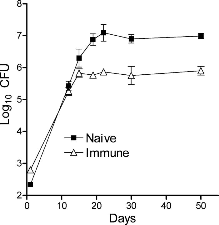
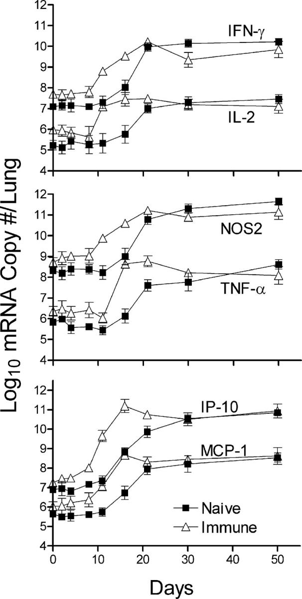
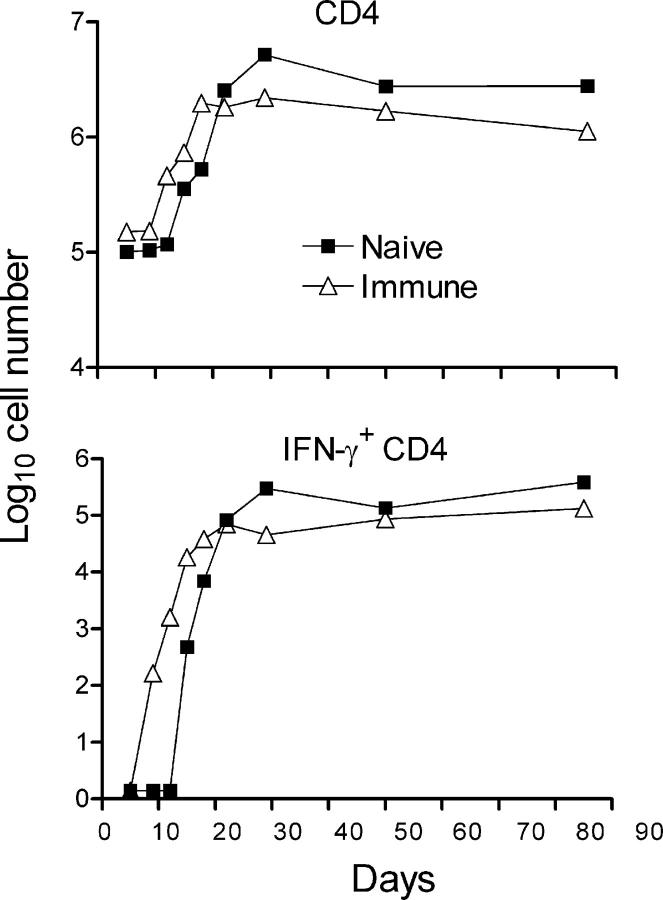
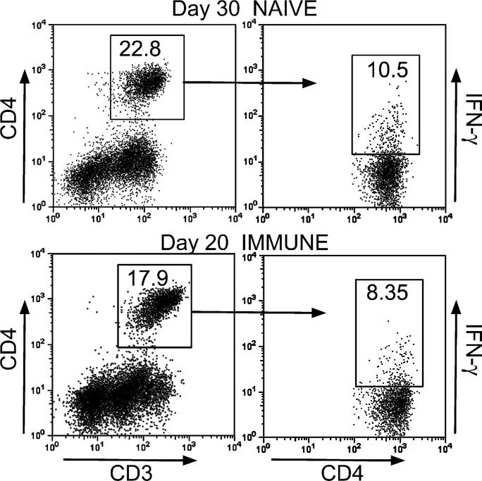
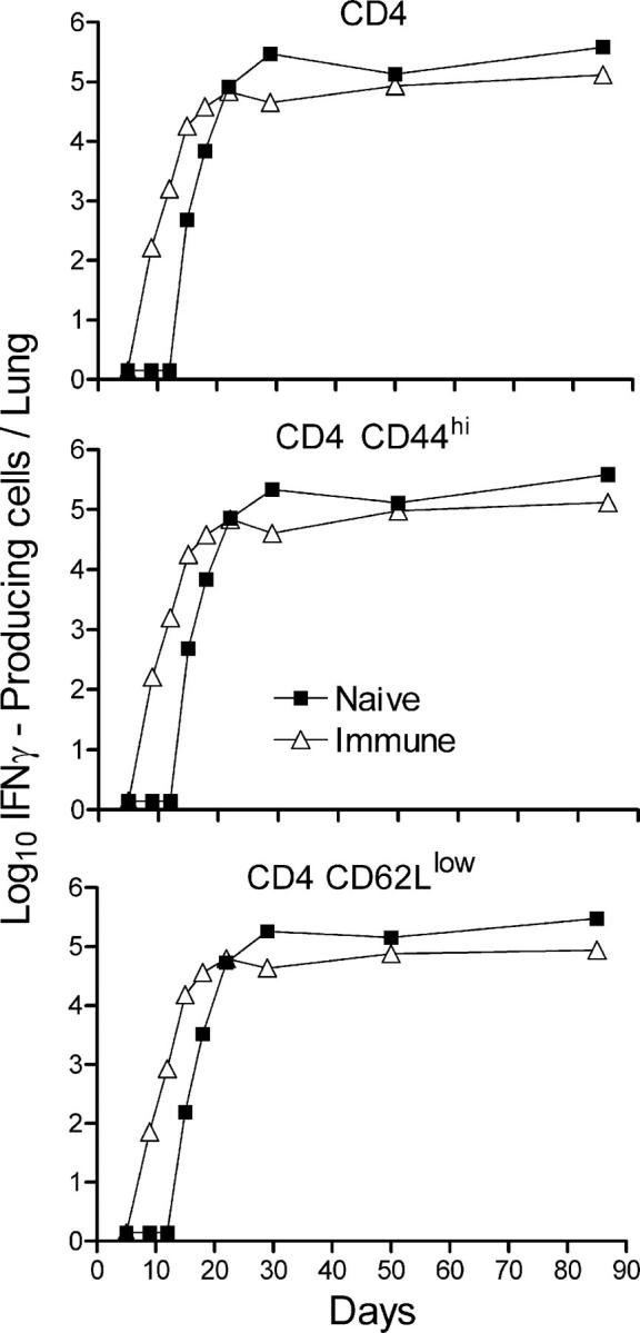
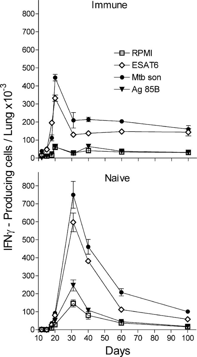
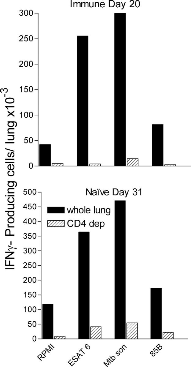
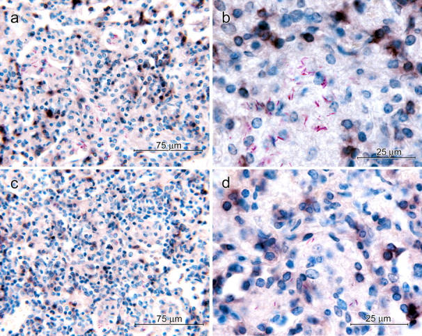
Mice immunized against Mycobacterium tuberculosis (Mtb) infection by curing them of a primary lung infection were compared with naive mice in terms of the ability to generate a Th1 cell immune response and to control growth of an airborne Mtb challenge infection. Immunized mice generated and expressed Th1 cell immunity several days sooner than naive mice, as demonstrated by an earlier increase in the synthesis in the lungs of mRNA for Th1 cytokines and for inducible nitric oxide synthase, an indicator of macrophage activation. This Th1 cytokine/mRNA synthesis was accompanied by an earlier accumulation of Mtb-specific Th1 cells in the lungs and the presence of CD4 T cells in lesions. An earlier generation of immunity was associated with an earlier inhibition of Mtb growth when infection was at a 1-log lower level. However, inhibition of Mtb growth in immunized, as well as in naive, mice was not followed by resolution of the infection, but by stabilization of the infection at a stationary level. The results indicate that there is no reason to believe that the secondary response to an Mtb infection is quantitatively or qualitatively superior to the primary response.


Figures

Figure 1.
Figure 1.
Course of Mtb infection in the lungs of immunized and naive mice infected with ∼2 × 10² CFUs of Mtb via the airborne route. Mtb grew log linearly for ∼20 d in the lungs of naive mice and for 15 d in the lungs of immunized mice before further growth was inhibited. Both in naive and immunized mice, inhibition of Mtb growth was followed by a stationary level infection that was 1 log lower in immunized mice. Means ± SD of five mice per group.
Figure 2.
Figure 2.
Real-time RT-PCR quantification of changes in Th1 cytokine gene transcription in the lungs of naive and immunized mice. Large increases in the synthesis of mRNA for IL-2, IFN-γ, and TNF-α were evident by day 12 of infection in immunized mice, 4 d earlier than in naive mice. The same applied to the increased synthesis of mRNA for IP-10 and MCP-10, and for NOS2. After day 12 in immunized and day 16 in naive mice, mRNA synthesis continued to increase to peak at 1–3 log higher levels, depending on the particular mRNA. Peak levels of synthesis were reached earlier in immunized mice, but were no higher than in naive mice. In all cases, mRNA synthesis was sustained at high levels until day 50, when the experiment was terminated. The results were obtained with the pooled lung RNA of four mice per time point per group. Shown are the means and SDs of four separate readings. The experiment was performed twice with essentially the same result.
Figure 3.
Figure 3.
The results of a flow cytometry analysis of the number of CD4 T cells and IFN-γ–secreting CD4 T cells that accumulated in the lungs of naive and immunized mice against time of the challenge infection. CD4 T cells began accumulating earlier in immunized mice and reached peak numbers on day 20, after which the number remained relatively constant. In naive mice, the peak number was reached on day 30. The same kinetics applied to the accumulation of IFN-γ–producing CD4 T cells in the naive and immunized mice, except that only a small percentage of the CD4 cells were positive for IFN-γ. The results were obtained with pooled lung cells from four mice stained for surface CD4 and intracellular IFN-γ as described in Materials and methods. The same result was obtained in two separate experiments.
Figure 4.
Figure 4.
Only a small percentage of CD4 T cells were making IFN-γ at peak responses. Only 10.5% of CD4 T cells in the lungs of naive mice (day 30), and 8.35% in the lungs of immunized mice (day 20), were making IFN-γ according to flow cytometry. The results were obtained with pooled lung cells of four mice stained for surface CD3 and CD4 and for intracellular IFN-γ as described in Materials and methods.
Figure 5.
Figure 5.
IFN-γ–producing CD4 T cells in the lungs displayed an activation phenotype. The majority of the CD4 T cells making IFN-γ in naive and vaccinated mice in response to a challenge infection displayed the CD44hiCD62Llow activation surface phenotype. Cells were stained for surface CD4, CD62L, and CD44, and intracellular IFN-γ as described in Materials and methods.
Figure 6.
Figure 6.
Kinetics of accumulation of Mtb-specific T cells in the lungs of naive and immunized mice. T cells capable of synthesizing IFN-γ in response to exposure to ESAT-61-20 peptide, Ag85240-254 peptide, and Mtb sonicate were enumerated with the Elispot assay. Increased numbers of Mtb-specific T cells were present in the lungs of immunized mice by day 12 of infection, but not until day 18 in the lungs of naive mice. Mtb-specific cells increased to peak on day 21 in the case of immunized mice and on day 30 in the case of naive mice. Consequently, Mtb-specific cells accumulated in higher numbers in the lungs of the latter mice. Results were obtained with pooled lung cells from four mice. Shown are the means ± SD of the number of spots in triplicate wells. The same result was obtained in two separate experiments.
Figure 7.
Figure 7.
Evidence that the majority of cells that made IFN-γ in the Elispot assay were CD4 positive. Magnetic depletion of ≥90% CD4 T cells from the lung cell suspension resulted in the removal of the majority of cells capable of making IFN-γ in response to ESAT-61-20 peptide, to Ag85B240-254 peptide, or to the Mtb sonicate in the Elispot assay. The experiment was performed with pooled cells harvested from four immunized mice on day 20 and from four naive mice on day 31 of infection.
Figure 8.
Figure 8.
Immunocytochemical demonstration of CD4 cells in lung lesions of immunized and naive mice at the peak of CD4 cell accumulation. 40× micrographs of lung lesion of a 30-d-infected naive mouse (a) and a 21-d-infected immunized mouse (c) showing CD4-positive cells (dark brown) throughout the lesion. 100× micrographs of a lesion of a naive (b) and immunized mouse (d) showing some CD4-positive cells in proximity to macrophages containing acid-fast bacilli. Discrete black/brown smudges represent CD4-positive cells out of the plane of focus.

Similar articles

Cited by

References

    1. Corbett, E.L., C.J. Watt, N. Walker, D. Maher, B.G. Williams, M.C. Raviglione, and C. Dye. 2003. The growing burden of tuberculosis: global trends and interactions with the HIV epidemic. Arch. Intern. Med. 163:1009–1021. - PubMed
    1. Orme, I.M. 1999. Beyond BCG: the potential for a more effective TB vaccine. Mol. Med. Today. 5:487–492. - PubMed
    1. Flynn, J.L., and J. Chan. 2001. Immunology of tuberculosis. Annu. Rev. Immunol. 19:93–129. - PubMed
    1. North, R.J., and Y.J. Jung. 2004. Immunity to tuberculosis. Annu. Rev. Immunol. 22:599–623. - PubMed
    1. Farer, L.S., A.M. Lowell, and W.B. Meador. 1979. Extrapulmonary tuberculosis in the United States. Am. J. Epidemiol. 109:205–217. - PubMed

Publication types

MeSH terms