. 2005 Jun 20;169(6):909-20.
doi: 10.1083/jcb.200501126. Epub 2005 Jun 13.

A positive feedback loop between Dumbfounded and Rolling pebbles leads to myotube enlargement in Drosophila

Affiliations

A positive feedback loop between Dumbfounded and Rolling pebbles leads to myotube enlargement in Drosophila

Sree Devi Menon et al. J Cell Biol. .

Abstract

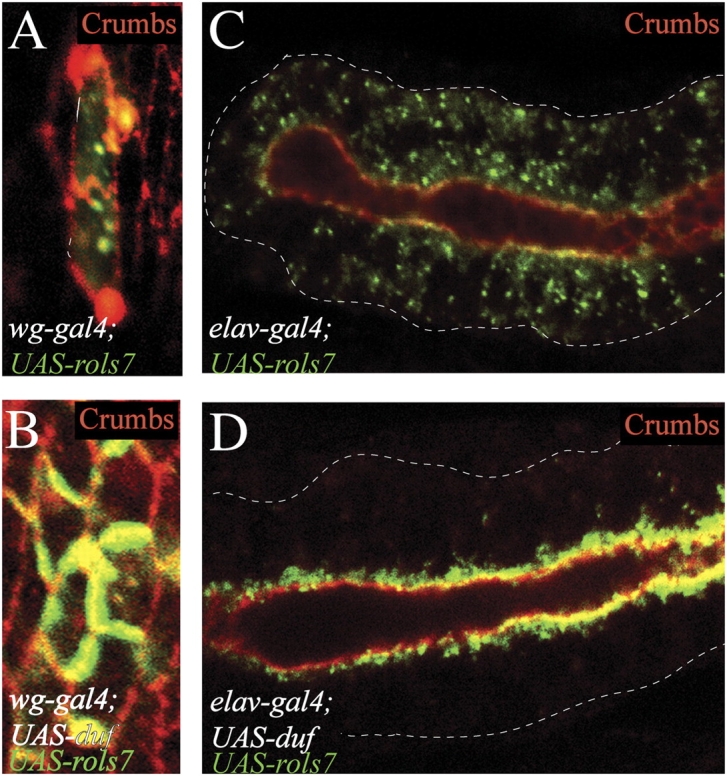
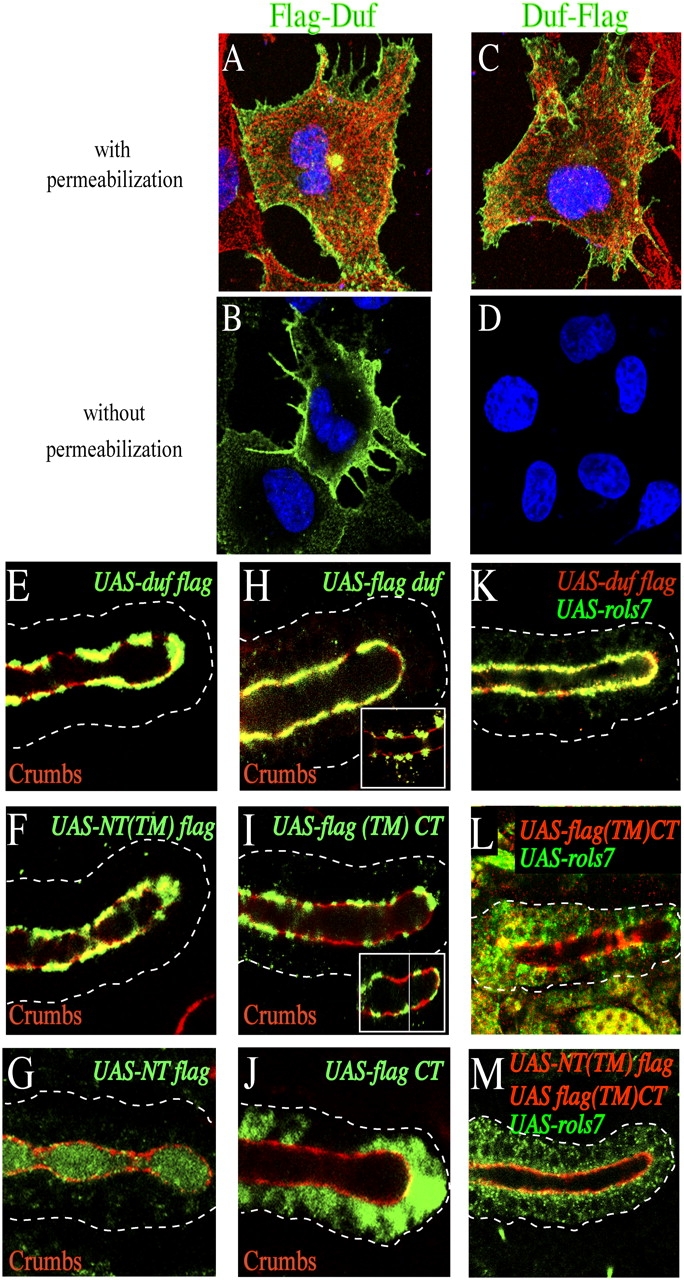
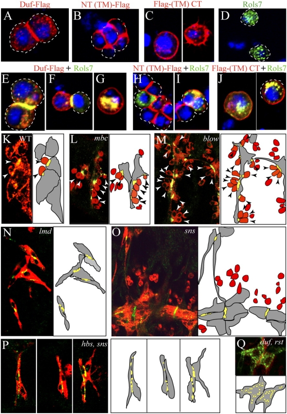
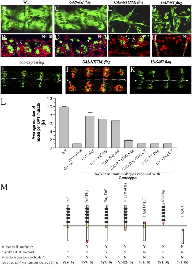
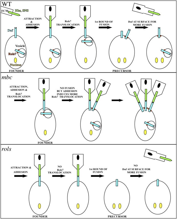
In Drosophila, myoblasts are subdivided into founders and fusion-competent myoblasts (fcm) with myotubes forming through fusion of one founder and several fcm. Duf and rolling pebbles 7 (Rols7; also known as antisocial) are expressed in founders, whereas sticks and stones (SNS) is present in fcm. Duf attracts fcm toward founders and also causes translocation of Rols7 from the cytoplasm to the fusion site. We show that Duf is a type 1 transmembrane protein that induces Rols7 translocation specifically when present intact and engaged in homophilic or Duf-SNS adhesion. Although its membrane-anchored extracellular domain functions as an attractant and is sufficient for the initial round of fusion, subsequent fusions require replenishment of Duf through cotranslocation with Rols7 tetratricopeptide repeat/coiled-coil domain-containing vesicles to the founder/myotube surface, causing both Duf and Rols7 to be at fusion sites between founders/myotubes and fcm. This implicates the Duf-Rols7 positive feedback loop to the occurrence of fusion at specific sites along the membrane and provides a mechanism by which the rate of fusion is controlled.


Figures

Figure 1.
Figure 1.
Rols7 translocates to adherence junctions in polarized nonmesodermal tissues when Duf is present. Rols7 was expressed in epidermal (A and B) or salivary gland (C and D) epithelium using the gal4-UAS system. Embryos were stained with antibodies against Rols7 (green) and Crumbs, a marker for adherence junctions (red). Dashes outline epidermal cell or salivary gland.
Figure 2.
Figure 2.
The intact Duf type 1 TM protein induces Rols7 to translocate. (A–D) Duf localization and topology. Full-length Duf Flag-tagged at its NH2 (A and B) or COOH terminus (C and D) was expressed in Cos cells. Cells were stained with anti-Flag antibodies (green), anti-tubulin antibodies (red), and Hoechst (blue). (E–J) Full-length or truncated Duf was expressed in the salivary gland and detected using antibodies against Flag (green, constructs shown schematically in Fig. 4 M). Crumbs marks adherence junctions (red). (K–M) Coexpression of Flag-tagged Duf constructs (red) and Rols7 (green) in the salivary gland. NT, NH2-terminal/EC; CT, COOH-terminal/IC. Position of tag in construct indicated by where “Flag” is placed in nomenclature. Dashes outline salivary gland.
Figure 3.
Figure 3.
Engagement of the Duf receptor through homophilic or Duf–SNS adhesion initiates and directs translocation of Rols7. (A–J) Full-length Duf, membrane-anchored Duf EC or IC regions (red) were expressed alone or together with Rols7 (green) in S2 cells. Dashes outline cell surface. Hoechst-labeled DNA in blue. (K–Q) WT (early stage 15) and mutant embryos (stage 16) were stained with anti-Rols7 (green) and anti-myosin heavy chain (MHC, red) to determine Rols7 distribution in relation to myoblast adhesion. Schematics show founders/myotubes in gray, fcm in red, and Rols7 in green (or yellow when it overlaps with MHC); arrowheads highlight fcm that adhere to founders/myotubes.
Figure 4.
Figure 4.
Functional analyses of Duf. (A–H) Full-length and Duf deletions were introduced into duf, rst mutants using the 24B-gal4 driver. Embryos were stained with anti-MHC antibodies (A, C, E, and G, showing dorsal muscles) or double labeled with antibodies against Eve (green nuclear stain) and either D-Titin (B, red cytoplasmic stain) or MHC (D, F, and H). Founders and myotubes outlined with dashes; arrowheads indicate adherent fcm. (I–K) Restoration of myoblast attraction as gauged by clustering of somatic fcm at epidermal sites from ectopic expression of constructs using the wingless (wg)-gal4 driver. Panels show ventral view of late stage 14 embryos with ventral midline demarcated by dashes. Anti-MHC stain (red) marks myoblasts and anti-Wg stain (green) marks the alternating strips of wg-gal4-driven expression domains in the epidermis. Mutant embryos expressing NT(TM)-Flag show fcm extending filopodia toward and adhering to the Wg-expressing epidermal regions (J) in contrast to nonexpressing or NT-Flag–expressing mutant embryos where fcm are not seen (I and K). (L) Extent of fusion in the somatic muscles was assessed based on the average number of Eve-positive nuclei within DA1 muscles at stage 15, denoted as “N” ± SD, from three independent experiments where 20–30 abdominal hemisegments from two to three embryos were analyzed at a time. A WT DA1 contains an average of 10 nuclei (n = 9.9 ± 0.3, whereas those in duf;rst Df(1)w67k30 mutants are mono-nucleated (n = 1.0 ± 0.0). Embryos were also stained with an antibody against Rols7 to ascertain its localization. Results are summarized in M. Yellow bar, TM region; orange oval, Flag tag; black circles, immunoglobulin repeats; Y (yes), N (no), Y/N (partial). As expression levels of Duf constructs in myoblasts was beyond the sensitivity of detection, its localization in M is as observed when constructs are ectopically expressed in the epidermis or salivary gland.
Figure 5.
Figure 5.
Distinct regions of Rols7 perform different functions. (A–F) HA-tagged full-length Rols7, deletions, and point mutations of catalytic residues in the putative lipolytic enzyme signature sequence were expressed alone or with Duf in the salivary gland. Glands were stained with an antibody against HA to ascertain Rols7 localization (green, constructs shown schematically in H). (G) Constructs were introduced into rols P1729ex18/Df(3L)BK9 mutant myoblasts using the 24B-gal4 driver and extent of fusion assessed by the average number of Eve-positive nuclei, “N” ± SD, within a DA1 muscle at stage 15 in comparison to 9.9 ± 0.3 in WT and 3.3 ± 0.2 in rols mutants. Data represent results from three independent experiments in which 20–30 abdominal hemisegments from two to three embryos were analyzed each time. (H) Summary of data obtained in G. Rols7 subcellular distribution was ascertained as described before. L (red), putative lipolytic enzyme sequence; R (orange), RING finger; U (white box), a region in Rols7 that is absent in Rols6; A (blue), ankyrin repeats; T (green), TPR repeats; black dash, coled-coil region; yellow oval, HA tag; Y (yes), N (no), Y/N (partial); nd, not detected. Position of tag in construct indicated by where “HA” is placed in nomenclature.
Figure 6.
Figure 6.
Rols7 sustains Duf expression and allows fusion to progress. (A–F) WT and mutant embryos were double labeled with an antibody raised against the Duf EC region (green) and another against MHC (red). Schematics depict founders/myotubes in gray, fcm in red and Duf in green (or yellow where it overlaps with MHC); arrowheads show adherent fcm. (G and H) Embryos carrying the D-mef2 mutation alone (G) or in combination with the rols mutation (H) were stained with antibodies raised against Eve (red nuclear stain), Rols7 (red cytoplasmic stain), and the Duf IC region (green cytoplasmic stain). Dashes outline founders. (I–Q) Indirect immunofluorescent analyses of ectopically expressed epitope-tagged Duf and Rols7. (I–K) Actively fusing dorsal myotubes at stage 13/14 with DA1 labeled with anti-Eve (blue nuclei). (L–N) Ventral myotubes at stage 13/14. (O–Q) After fusion ventral muscles at stage 16. Schematics show myotubes/muscles in gray and overlapping Duf and Rols7 expression in yellow. When ectopically expressed in actively fusing myotubes, both Duf and Rols7 are visible as cytoplasmic puncta and rarely seen as “dashes” or “lines” at the myotube surface (L and N, arrowheads). At stage 16, both proteins clearly localize at the surface of the ventral muscles, particularly at muscle-muscle adhesion sites. (R) The extent of fusion in various mutants gauged by the average number of Eve-positive nuclei “N” ± SD within a DA1 muscle at stage 15. Data represent results from three independent experiments in which 20–30 abdominal hemisegments from two to three embryos were analyzed at a time.
Figure 7.
Figure 7.
Schematic of events envisaged to occur in WT and mutant myoblasts. (Top) Duf localizes at the WT founder cell surface. Its EC region serves as an attractant, binds Hbs and SNS and, in doing so, brings about adhesion between founder and fcm. The first round of intercellular fusion ensues, culminating in the formation of a precursor, and this may result in the cleavage/removal of the Duf receptor from the cell surface. At the same time, adhesion between the founder and fcm initiates IC events in the founder that lead to translocation (and possibly fusion) of Duf-loaded Rols7 vesicles to the adhesion site. The new batch of Duf at the surface initiates another round of myoblast fusion. (Middle) In mbc or D-mef2 mutants, engagement of Duf induces Rols7 vesicles to translocate and deposit the next batch of Duf at the founder cell surface. However, in these mutants, fusion does not occur and the persistent adhesion between fcm and the founder leads to additional Rols7 and Duf being deposited at the founder surface. In lmd and sns mutants, similar events triggered by homophilic Duf adhesion causes Rols7 and Duf to be targeted to sites of contact between founders. (Bottom) In the rols mutant, the first round of fusion occurs undisrupted. However, without Rols7, Duf levels at the precursor surface is not replenished and fusion stalls. Surface levels of Rst may be similar regulated.

Similar articles

Cited by

References

    1. Abmayr, S.M., L. Balagopalan, B.J. Galletta, and S.J. Hong. 2003. Cell and molecular biology of myoblast fusion. Int. Rev. Cytol. 225:33–89. - PubMed
    1. Artero, R.D., I. Castanon, and M.K. Baylies. 2001. The immunoglobulin-like protein Hibris functions as a dose-dependent regulator of myoblast fusion and is differentially controlled by Ras and Notch signaling. Development. 128:4251–4264. - PubMed
    1. Bate, M. 1990. The embryonic development of larval muscles in Drosophila. Development. 110:791–804. - PubMed
    1. Baylies, M.K., M. Bate, and G.M. Ruiz. 1998. Myogenesis: a view from Drosophila. Cell. 93:921–927. - PubMed
    1. Bour, B.A., M.A. O'Brien, W.L. Lockwood, E.S. Goldstein, R. Bodmer, P.H. Taghert, S.M. Abmayr, and H.T. Nguyen. 1995. Drosophila MEF2, a transcription factor that is essential for myogenesis. Genes Dev. 9:730–741. - PubMed

Publication types

MeSH terms