

Loss of G-A base pairs is insufficient for achieving a large opening of U4 snRNA K-turn motif
- PMID: 15956103
- PMCID: PMC1150281
- DOI: 10.1093/nar/gki664
Loss of G-A base pairs is insufficient for achieving a large opening of U4 snRNA K-turn motif
Abstract
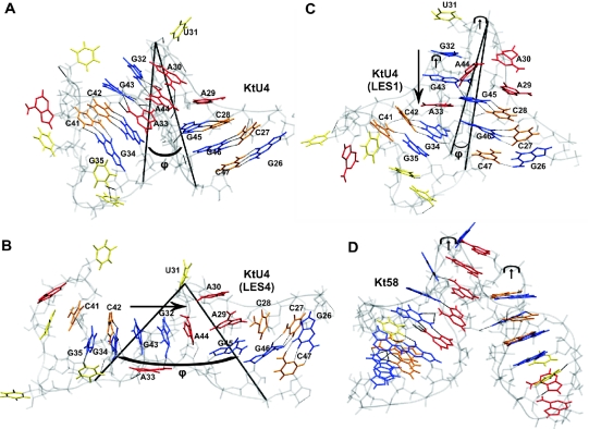
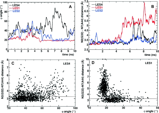
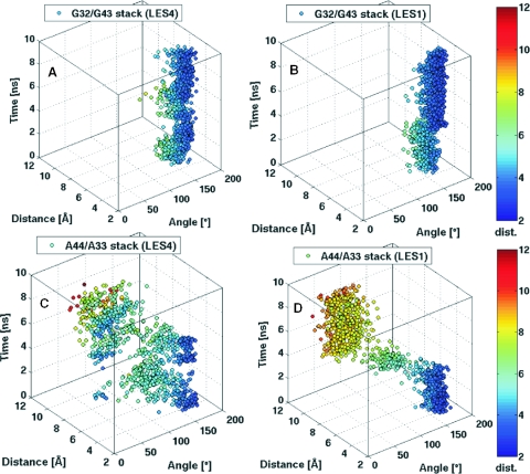
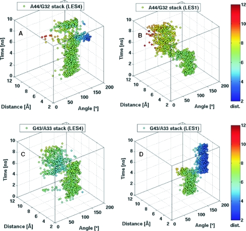
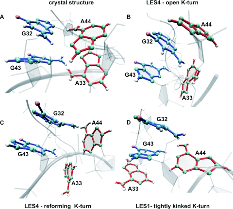
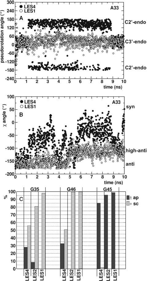
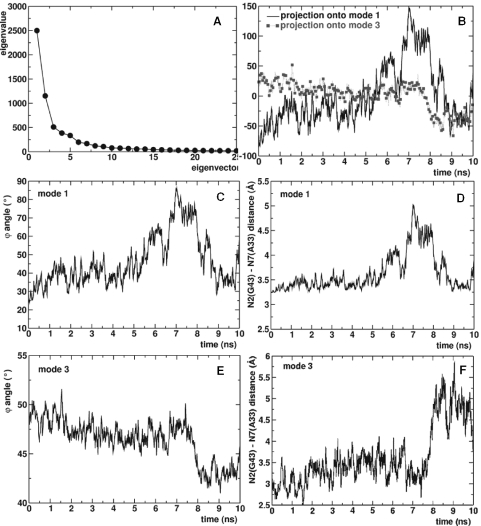
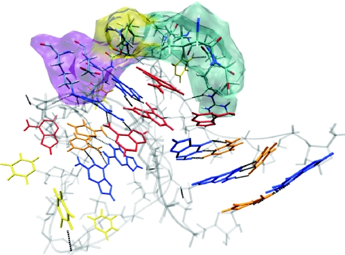
Upon binding to the 15.5K protein, two tandem-sheared G-A base pairs are formed in the internal loop of the kink-turn motif of U4 snRNA (Kt-U4). We have reported that the folding of Kt-U4 is assisted by protein binding. Unstable interactions that contribute to a large opening of the free RNA ('k-e motion') were identified using locally enhanced sampling molecular dynamics simulations, results that agree with experiments. A detailed analysis of the simulations reveals that the k-e motion in Kt-U4 is triggered both by loss of G-A base pairs in the internal loop and backbone flexibility in the stems. Essential dynamics show that the loss of G-A base pairs is correlated along the first mode but anti-correlated along the third mode with the k-e motion. Moreover, when enhanced sampling was confined to the internal loop, the RNA adopted an alternative conformation characterized by a sharper kink, opening of G-A base pairs and modified stacking interactions. Thus, loss of G-A base pairs is insufficient for achieving a large opening of the free RNA. These findings, supported by previously published RNA structure probing experiments, suggest that G-A base pair formation occurs upon protein binding, thereby stabilizing a selective orientation of the stems.
Figures
Similar articles
-
Detecting protein-induced folding of the U4 snRNA kink-turn by single-molecule multiparameter FRET measurements.RNA. 2005 Oct;11(10):1545-54. doi: 10.1261/rna.2950605. RNA. 2005. PMID: 16199764 Free PMC article.
-
RNA structure and RNA-protein interactions in purified yeast U6 snRNPs.J Mol Biol. 2006 Mar 10;356(5):1248-62. doi: 10.1016/j.jmb.2005.12.013. Epub 2005 Dec 20. J Mol Biol. 2006. PMID: 16410014
-
The snRNP 15.5K protein folds its cognate K-turn RNA: a combined theoretical and biochemical study.RNA. 2005 Feb;11(2):197-209. doi: 10.1261/rna.7149605. RNA. 2005. PMID: 15659359 Free PMC article.
-
Structural dynamics of the box C/D RNA kink-turn and its complex with proteins: the role of the A-minor 0 interaction, long-residency water bridges, and structural ion-binding sites revealed by molecular simulations.J Phys Chem B. 2010 Aug 19;114(32):10581-93. doi: 10.1021/jp102572k. J Phys Chem B. 2010. PMID: 20701388
-
The kink-turn in the structural biology of RNA.Q Rev Biophys. 2018 Jan;51:e5. doi: 10.1017/S0033583518000033. Q Rev Biophys. 2018. PMID: 30912490 Review.
Cited by
-
Identification of compound heterozygous variants in the noncoding RNU4ATAC gene in a Chinese family with two successive foetuses with severe microcephaly.Hum Genomics. 2018 Jan 25;12(1):3. doi: 10.1186/s40246-018-0135-9. Hum Genomics. 2018. PMID: 29370840 Free PMC article.
-
RNA Structural Dynamics As Captured by Molecular Simulations: A Comprehensive Overview.Chem Rev. 2018 Apr 25;118(8):4177-4338. doi: 10.1021/acs.chemrev.7b00427. Epub 2018 Jan 3. Chem Rev. 2018. PMID: 29297679 Free PMC article. Review.
-
RNA kink-turns are highly anisotropic with respect to lateral displacement of the flanking stems.Biophys J. 2022 Mar 1;121(5):705-714. doi: 10.1016/j.bpj.2022.01.025. Epub 2022 Feb 3. Biophys J. 2022. PMID: 35122735 Free PMC article.
-
The role of specific 2'-hydroxyl groups in the stabilization of the folded conformation of kink-turn RNA.RNA. 2007 Feb;13(2):200-10. doi: 10.1261/rna.285707. Epub 2006 Dec 8. RNA. 2007. PMID: 17158708 Free PMC article.
-
Elastic properties of ribosomal RNA building blocks: molecular dynamics of the GTPase-associated center rRNA.Nucleic Acids Res. 2007;35(12):4007-17. doi: 10.1093/nar/gkm245. Epub 2007 Jun 6. Nucleic Acids Res. 2007. PMID: 17553840 Free PMC article.
References
-
- Vidovic I., Nottrott S., Hartmuth K., Lührmann R., Ficner R. Crystal structure of the spliceosomal 15.5kD protein bound to a U4 snRNA fragment. Mol. Cell. 2000;6:1331–1342. - PubMed
-
- Charron C., Manival X., Clery A., Charpentier B., Marmier-Gourrier N., Branlant C., Aubry A. The archaeal sRNA binding protein L7Ae has a 3D structure very similar to that of its eukaryal counterpart while having a broader RNA-binding specificity. J. Mol. Biol. 2004;342:757–773. - PubMed
