. 2005 Jun 21;102(25):8893-8.
doi: 10.1073/pnas.0503750102. Epub 2005 Jun 14.

NMR study of a membrane protein in detergent-free aqueous solution

Affiliations

NMR study of a membrane protein in detergent-free aqueous solution

Manuela Zoonens et al. Proc Natl Acad Sci U S A. .

Abstract

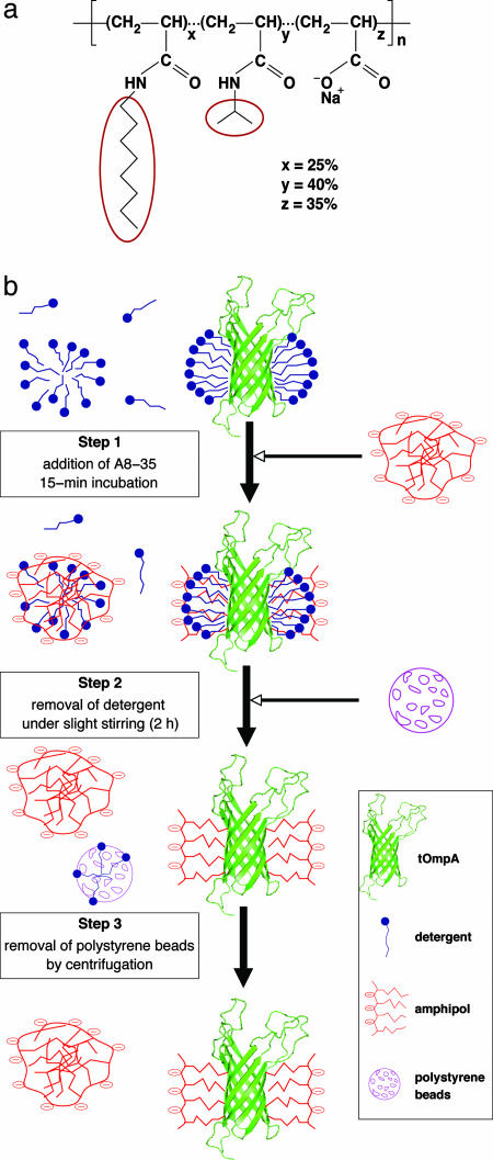
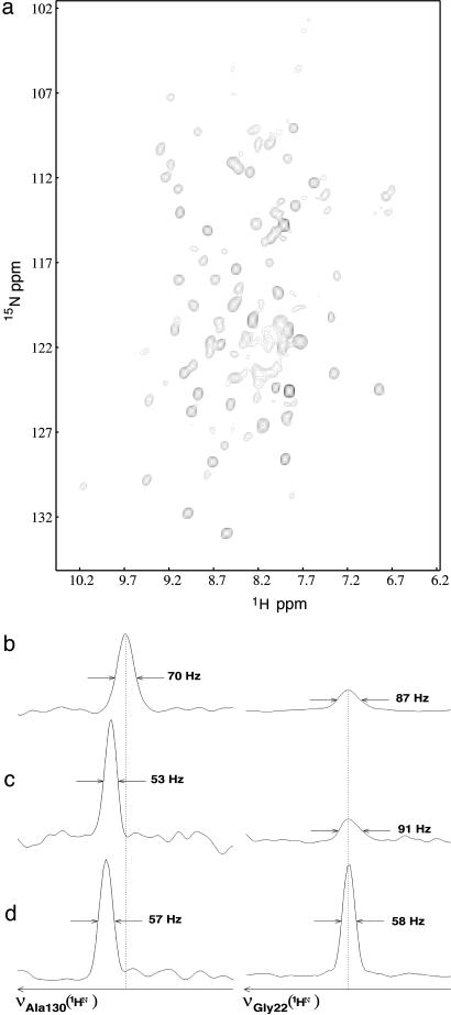
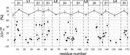
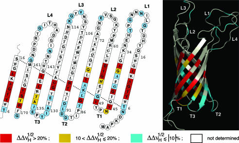
One of the major obstacles to membrane protein (MP) structural studies is the destabilizing effect of detergents. Amphipols (APols) are short amphipathic polymers that can substitute for detergents to keep MPs water-soluble under mild conditions. In the present work, we have explored the feasibility of studying the structure of APol-complexed MPs by NMR. As a test MP, we chose the 171-residue transmembrane domain of outer MP A from Escherichia coli (tOmpA), whose x-ray and NMR structures in detergent are known. 2H,15N-labeled tOmpA was produced as inclusion bodies, refolded in detergent solution, trapped with APol A8-35, and the detergent removed by adsorption onto polystyrene beads. The resolution of transverse relaxation-optimized spectroscopy-heteronuclear single-quantum correlation spectra of tOmpA/A8-35 complexes was found to be close to that of the best spectra obtained in detergent solutions. The dispersion of chemical shifts indicated that the protein had regained its native fold and retained it during the exchange of surfactants. MP-APol interactions were mapped by substituting hydrogenated for deuterated A8-35. The resulting dipolar broadening of amide proton linewidths was found to be limited to the beta-barrel region of tOmpA, indicating that A8-35 binds specifically to the hydrophobic transmembrane surface of the protein. The potential of this approach to MP studies by solution NMR is discussed.


Figures

Fig. 1.
Fig. 1.
Trapping tOmpA with amphipol A8-35. (a) Chemical structure of A8-35, a polyacrylate-based APol. The average degree of polymerization (n) is ≈80, and the average molecular weight, ≈9 kDa. The molar percentage of each type of unit (x, y, and z), randomly distributed along the chain, is indicated. It corresponds, respectively, to ≈20, ≈32, and ≈28 units per A8-35 molecule. HAPol is the hydrogenated form; in DAPol, the octyl and isopropyl side chains are perdeuterated (ellipses). (b) Trapping procedure; see text.
Fig. 2.
Fig. 2.
NMR 2D spectrum of A8-35-trapped tOmpA. (a) [1H,15N]-TROSY-HSQC spectrum of [2H,15N]tOmpA/HAPol recorded at 30°C, pH 7.9, and 800 MHz 1H frequency. (b) Zooms on two 1H rows, one showing Ala-130, a residue belonging to the β-barrel (Left), the other Gly-22, a residue from an extracellular loop (Right), extracted from the spectrum shown in a.(c and d) Signals of the same residues obtained with a tOmpA/DHPC sample at pH 7.9 and 600 MHz (c) and pH 6.5 and 800 MHz (d). TROSY spectra were aligned relative to each other by taking the 1HN-15N line of Gly-22 as an internal reference.
Fig. 3.
Fig. 3.
Dipolar broadening due to tOmpA/APol interactions. Changes in 1HN linewidth variations, measured at midheight (formula image), for tOmpA trapped with a fully protonated APol (HAPol) with respect to signals obtained after complexation with an APol with perdeuterated alkyl chains (DAPol) are plotted against residue number. 2D TROSY experiments were repeated at least three times. Average variations are shown ± SD. Squares, diamonds, and circles refer to amino acids belonging to β-strands, periplasmic turns, and external loops, respectively. Averages for each secondary structure element are shown at the top, under a schematic representation of the secondary structure.
Fig. 4.
Fig. 4.
Mapping of contacts with APol alkyl chains onto the structure of tOmpA. Residues are color-coded depending on whether dipolar line broadening upon substituting HAPol for DAPol (Fig. 3) is strong (red), weak (yellow), or undetectable (blue). Residues giving rise to no assigned line are white. The data are plotted on a topology sketch (adapted from ref. ; the side chains of residues shown in italics point toward the exterior of the barrel) and on a ribbon representation of the 3D structure (Protein Data Bank code 1G90; ref. 6). The 3D diagram was realized with the open-source software pymol (DeLano Scientific, San Carlos, CA).

References

    1. Liu, J. & Rost, B. (2001) Protein Sci. 10, 1970–1979. - PMC - PubMed
    1. le Maire, M., Champeil, P. & Møller, J. V. (2000) Biochim. Biophys. Acta 1508, 86–111. - PubMed
    1. Bowie, J. U. (2001) Curr. Opin. Struct. Biol. 11, 397–402. - PubMed
    1. McDonnell, P. A. & Opella, S. J. (1993) J. Magn. Reson. B102, 120–125.
    1. MacKenzie, K. R., Prestegard, J. H. & Engelman, D. M. (1997) Science 276, 131–133. - PubMed

Publication types