

Rapid plasticity of binocular connections in developing monkey visual cortex (V1)
- PMID: 15956191
- PMCID: PMC1150295
- DOI: 10.1073/pnas.0500280102
Rapid plasticity of binocular connections in developing monkey visual cortex (V1)
Abstract
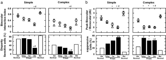
The basic sets of cortical connections are present at birth in the primate visual system. The maintenance and refinement of these innate connections are highly dependent on normal visual experience, and prolonged exposure to binocularly uncorrelated signals early in life severely disrupts the normal development of binocular functions. However, very little is known about how rapidly these changes in the functional organization of primate visual cortex emerge or what are the sequence and the nature of the abnormal neural events that occur immediately after experiencing binocular decorrelation. In this study, we investigated how brief periods of ocular misalignment (strabismus) at the height of the critical period alter the cortical circuits that support binocular vision. After only 3 days of optically imposed strabismus, there was a striking increase in the prevalence of V1 neurons that exhibited binocular suppression, i.e., binocular responses were weaker than monocular responses. However, the sensitivity of these neurons to interocular spatial phase disparity was not significantly altered. These contrasting results suggest that the first significant change in V1 caused by early binocular decorrelation is binocular suppression, and that this suppression originates at a site(s) beyond where binocular signals are initially combined.
Figures


Similar articles
-
Effects of the duration of early strabismus on the binocular responses of neurons in the monkey visual cortex (V1).Invest Ophthalmol Vis Sci. 2002 Apr;43(4):1262-9. Invest Ophthalmol Vis Sci. 2002. PMID: 11923274
-
Effect of onset age of strabismus on the binocular responses of neurons in the monkey visual cortex.Invest Ophthalmol Vis Sci. 2000 Mar;41(3):948-54. Invest Ophthalmol Vis Sci. 2000. PMID: 10711717
-
Residual binocular interactions in the striate cortex of monkeys reared with abnormal binocular vision.J Neurophysiol. 1997 Sep;78(3):1353-62. doi: 10.1152/jn.1997.78.3.1353. J Neurophysiol. 1997. PMID: 9310426
-
Visual callosal connections and strabismus.Behav Brain Res. 1994 Oct 20;64(1-2):85-95. doi: 10.1016/0166-4328(94)90121-x. Behav Brain Res. 1994. PMID: 7840895 Review.
-
The neural basis of suppression and amblyopia in strabismus.Eye (Lond). 1996;10 ( Pt 2):250-8. doi: 10.1038/eye.1996.54. Eye (Lond). 1996. PMID: 8776456 Review.
Cited by
-
Degraded attentional modulation of cortical neural populations in strabismic amblyopia.J Vis. 2016;16(3):16. doi: 10.1167/16.3.16. J Vis. 2016. PMID: 26885628 Free PMC article.
-
Observations on the relationship between anisometropia, amblyopia and strabismus.Vision Res. 2017 May;134:26-42. doi: 10.1016/j.visres.2017.03.004. Epub 2017 Apr 18. Vision Res. 2017. PMID: 28404522 Free PMC article. Review.
-
Leveraging neural plasticity for the treatment of amblyopia.Surv Ophthalmol. 2024 Sep-Oct;69(5):818-832. doi: 10.1016/j.survophthal.2024.04.006. Epub 2024 May 18. Surv Ophthalmol. 2024. PMID: 38763223 Free PMC article. Review.
-
V1 microcircuit dynamics: altered signal propagation suggests intracortical origins for adaptation in response to visual repetition.J Neurophysiol. 2019 May 1;121(5):1938-1952. doi: 10.1152/jn.00113.2019. Epub 2019 Mar 27. J Neurophysiol. 2019. PMID: 30917065 Free PMC article.
-
Local structural-functional connectivity decoupling of caudate nucleus in infantile esotropia.Front Neurosci. 2022 Dec 22;16:1098735. doi: 10.3389/fnins.2022.1098735. eCollection 2022. Front Neurosci. 2022. PMID: 36620443 Free PMC article.
References
-
- Kiorpes, L. & Movshon, J. A. (2003) in The Visual Neurosciences (MIT Press, Cambridge, MA), pp. 159–173.
-
- Chino, Y. M., Bi, H. & Zhang, B. (2004) in The Primate Visual System (CRC Press, Boca Raton, FL), pp. 81–108.
-
- Crawford, M. L. J. & von Noorden, G. K. (1979) Invest. Ophthalmol. Visual Sci. 18, 496–505. - PubMed
-
- Crawford, M. L. J. & von Noorden, G. K. (1980) Invest. Ophthalmol. Visual Sci. 20, 665–670. - PubMed
-
- Smith, E. L.. 3rd, Chino, Y. M., Ni, J., Cheng, H., Crawford, L. J. & Harwerth, S. (1997) J. Neurophysiol. 78, 1353–1362. - PubMed
Publication types
MeSH terms
Grants and funding
LinkOut - more resources
Full Text Sources
