. 2005 Jun 28;102(26):9247-52.
doi: 10.1073/pnas.0502040102. Epub 2005 Jun 13.

Evasion of Toll-like receptor 5 by flagellated bacteria

Affiliations

Evasion of Toll-like receptor 5 by flagellated bacteria

Erica Andersen-Nissen et al. Proc Natl Acad Sci U S A. .

Abstract

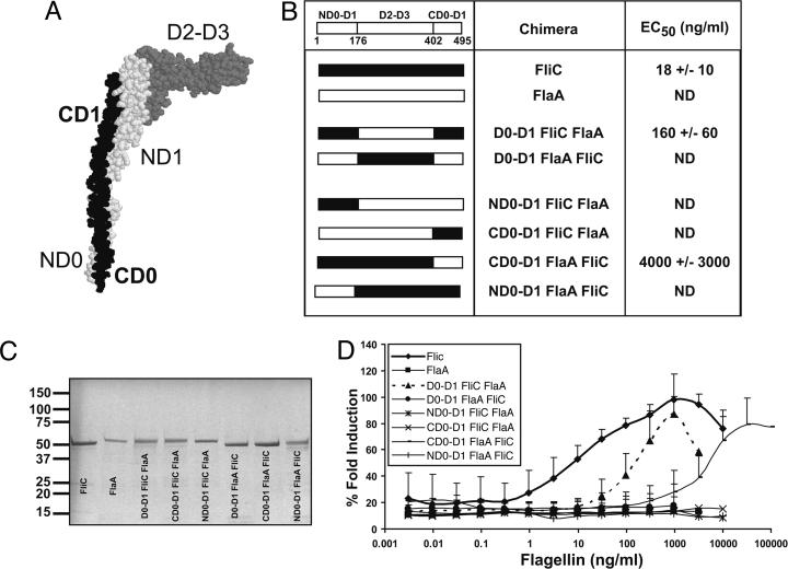
Toll-like receptor 5 (TLR5) recognizes an evolutionarily conserved site on bacterial flagellin that is required for flagellar filament assembly and motility. The alpha and epsilon Proteobacteria, including the important human pathogens Campylobacter jejuni, Helicobacter pylori, and Bartonella bacilliformis, require flagellar motility to efficiently infect mammalian hosts. In this study, we demonstrate that these bacteria make flagellin molecules that are not recognized by TLR5. We map the site responsible for TLR5 evasion to amino acids 89-96 of the N-terminal D1 domain, which is centrally positioned within the previously defined TLR5 recognition site. Salmonella flagellin is strongly recognized by TLR5, but mutating residues 89-96 to the corresponding H. pylori flaA sequence abolishes TLR5 recognition and also destroys bacterial motility. To preserve bacterial motility, alpha and epsilon Proteobacteria possess compensatory amino acid changes in other regions of the flagellin molecule, and we engineer a mutant form of Salmonella flagellin that evades TLR5 but retains motility. These results suggest that TLR5 evasion is critical for the survival of this subset of bacteria at mucosal sites in animals and raise the intriguing possibility that flagellin receptors provided the selective force to drive the evolution of these unique subclasses of bacterial flagellins.


Figures

Fig. 1.
Fig. 1.
Flagellin from α and ε Proteobacteria is not detected by TLR5. (A and B) Approximately 1 × 10⁵ CHO cells stably expressing human TLR5 and an NF-κB luciferase reporter were stimulated for 4 h with heat-killed samples of stationary phase bacterial cultures diluted 1:100 in media. Data represent % fold induction of luciferase activity relative to maximal stimulation achieved with Salmonella typhimurium (≈10- to 12-fold increase in luciferase activity over stimulation with LB alone) for at least three independent experiments, each run in triplicate. Error bars represent 1 SD. Control CHO cells stably transfected with empty expression vector and NF-κB luciferase reporter did not respond to flagellated bacteria (data not shown). (C) Immunoblots of sonicated samples of stationary phase bacterial cultures diluted 1:10. (C Left) Bartonella bacilliformis flagellin, as detected with anti-flagellin antiserum. (Center) Rhizobium (Ensifer) meliloti flagellin, as detected with anti-flagellin antiserum (bands at ≈23 and 26 kDa likely represent degraded flagellin). (Right) ε Proteobacteria flagellins as detected with anti-C. jejuni flagellin antiserum (additional band in H. pylori lanes likely represents crossreacting hook protein ≈75 kDa). The left margins show molecular size in kDa. (D) TLR5 dose-response curve to purified flagellins. Flagella were sheared from bacteria and purified by ultracentrifugation, and purity was confirmed by SDS/PAGE and Coomassie blue staining. Purified flagellin was incubated with CHO-hTLR5 cells for 4 h. ST, Salmonella typhimurium; EC, Escherichia coli; PA, Pseudomonas aeruginosa; LM, Listeria monocytogenes; HP, H. pylori; CJ, C. jejuni. Error bars represent 1 SD. (E) Molecular tree of flagellin sequences from flagellated bacteria, constructed by using clustalw and displayed with phylodendron. -, flagellated bacteria tested that did not activate TLR5; *, flagellated bacteria tested that activated TLR5.
Fig. 2.
Fig. 2.
The N-terminal D0-D1 domain of flagellin is required but not sufficient for TLR5 recognition. (A) Structure of FliC from Salmonella typhimurium with major domains labeled. The switch region (S) connects D0 and D1, but is not labeled. The protein Database (PDB) identification code 1ucu, is displayed in protein explorer. (B) Table listing flagellin chimeras made and the effective concentration required for 50% maximal TLR5 activation in ng/ml (EC50) ± SD. A linear schematic of FliC shows the amino acid numbers of the domain boundaries. ND, not detected. (C) Coomassie-stained SDS/PAGE of chimeric flagellins purified from Escherichia coli BL-21RIL (1 μg of protein loaded per lane). The left margin shows molecular size in kDa. (D) Dose-response curve of CHO-hTLR5 to purified flagellin chimeras as in 1 A.
Fig. 3.
Fig. 3.
Amino acids 89-96 are required for TLR5 activation. (A) Sequence alignment (clustalw) of flagellin proteins from bacteria that activate TLR5 with those of the α and ε Proteobacteria. Asterisks indicate residues in this region previously determined to be important for TLR5 recognition (3). (B) Table listing flagellin chimeras made and their corresponding EC50 ± SD. ND, not detected. (C) Dose-response curve of CHO-hTLR5 to purified flagellin chimeras. (D) Salmonella typhimurium BC696 (SL1344 fliC-fljB-) expressing wild-type or the 89-96 FlaA FliC flagellin chimera was stab-inoculated into motility agar and photographed. Data are representative of three independent experiments.
Fig. 4.
Fig. 4.
ε Proteobacteria possess compensatory changes that allow filament formation and motility. (A) Stacking of FliC monomers using PDB file 1io1 was performed according to Samatey et al. (24) and is represented in protein explorer to show contact surfaces. (A Left) View of two monomers: dark blue, concave contact surface; green and yellow, convex contact surface; yellow, amino acids 89-96. (A Right) Close-up of contact surface showing the location of 89-96 (yellow), I411 (red), and K58, G59 (light blue). Amino acids N87, L88, and R118 are represented as sticks to show the buried residue, I411. (B) Table listing mutant flagellins made and their corresponding EC50 ± SD. (C) Dose-response curves of CHO-hTLR5 stimulated with purified mutant flagellins. (D) Salmonella typhimurium BC696 (SL1344 fliC-fljB-) expressing wild-type or FliC mutant constructs were stab-inoculated into motility agar and photographed. Data are representative of three independent experiments.
Fig. 5.
Fig. 5.
Some members of the α Proteobacteria are predicted to be recognized by TLR5. Sequence alignment (clustalw) of flagellin proteins from bacteria that activate TLR5 with those of the α and ε Proteobacteria in the 89-96 region. Asterisks indicate residues in this region previously determined to be important for TLR5 recognition (3).

References

    1. Janeway, C. A., Jr., & Medzhitov, R. (2002) Annu. Rev. Immunol. 20, 197-216. - PubMed
    1. Medzhitov, R. & Janeway, C. A., Jr. (1997) Cell 91, 295-298. - PubMed
    1. Smith, K. D., Andersen-Nissen, E., Hayashi, F., Strobe, K., Bergman, M. A., Barrett, S. L., Cookson, B. T. & Aderem, A. (2003) Nat. Immunol. 4, 1247-1253. - PubMed
    1. Smith, M. F., Jr., Mitchell, A., Li, G., Ding, S., Fitzmaurice, A. M., Ryan, K., Crowe, S. & Goldberg, J. B. (2003) J. Biol. Chem. 278, 32552-32560. - PubMed
    1. Torok, A. M., Bouton, A. H. & Goldberg, J. B. (2005) Infect. Immun. 73, 1523-1531. - PMC - PubMed

Publication types

MeSH terms