The molecular basis of ferroportin-linked hemochromatosis
- PMID: 15956209
- PMCID: PMC1157058
- DOI: 10.1073/pnas.0503804102
The molecular basis of ferroportin-linked hemochromatosis
Abstract
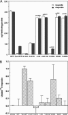
Mutations in the iron exporter ferroportin (Fpn) (IREG1, SLC40A1, and MTP1) result in hemochromatosis type IV, a disorder with a dominant genetic pattern of inheritance and heterogeneous clinical presentation. Most patients develop iron loading of Kupffer cells with relatively low saturation of plasma transferrin, but others present with high transferrin saturation and iron-loaded hepatocytes. We show that known human mutations introduced into mouse Fpn-GFP generate proteins that either are defective in cell surface localization or have a decreased ability to be internalized and degraded in response to hepcidin. Studies using co-immunoprecipitation of epitope-tagged Fpn and size-exclusion chromatography demonstrated that Fpn is multimeric. Both WT and mutant Fpn participate in the multimer, and mutant Fpn can affect the localization of WT Fpn, its stability, and its response to hepcidin. The behavior of mutant Fpn in cell culture and the ability of mutant Fpn to act as a dominant negative explain the dominant inheritance of the disease as well as the different patient phenotypes.
Figures


Similar articles
-
Resistance to hepcidin is conferred by hemochromatosis-associated mutations of ferroportin.Blood. 2005 Aug 1;106(3):1092-7. doi: 10.1182/blood-2005-02-0561. Epub 2005 Apr 14. Blood. 2005. PMID: 15831700
-
The molecular basis of hepcidin-resistant hereditary hemochromatosis.Blood. 2009 Jul 9;114(2):437-43. doi: 10.1182/blood-2008-03-146134. Epub 2009 Apr 21. Blood. 2009. PMID: 19383972 Free PMC article.
-
The flatiron mutation in mouse ferroportin acts as a dominant negative to cause ferroportin disease.Blood. 2007 May 15;109(10):4174-80. doi: 10.1182/blood-2007-01-066068. Epub 2007 Feb 8. Blood. 2007. PMID: 17289807 Free PMC article.
-
Non-HFE hepatic iron overload.Semin Liver Dis. 2011 Aug;31(3):302-18. doi: 10.1055/s-0031-1286061. Epub 2011 Sep 7. Semin Liver Dis. 2011. PMID: 21901660 Review.
-
Iron overload due to mutations in ferroportin.Haematologica. 2006 Jan;91(1):92-5. Haematologica. 2006. PMID: 16434376 Free PMC article. Review.
Cited by
-
Hamp1 but not Hamp2 regulates ferroportin in fish with two functionally distinct hepcidin types.Sci Rep. 2017 Nov 1;7(1):14793. doi: 10.1038/s41598-017-14933-5. Sci Rep. 2017. PMID: 29093559 Free PMC article.
-
Molecular and clinical correlates in iron overload associated with mutations in ferroportin.Haematologica. 2006 Aug;91(8):1092-5. Haematologica. 2006. PMID: 16885049 Free PMC article.
-
Iron imports. III. Transfer of iron from the mucosa into circulation.Am J Physiol Gastrointest Liver Physiol. 2006 Jan;290(1):G1-6. doi: 10.1152/ajpgi.00415.2005. Am J Physiol Gastrointest Liver Physiol. 2006. PMID: 16354771 Free PMC article. Review.
-
Understanding the structure/activity relationships of the iron regulatory peptide hepcidin.Chem Biol. 2011 Mar 25;18(3):336-43. doi: 10.1016/j.chembiol.2010.12.009. Chem Biol. 2011. PMID: 21439478 Free PMC article.
-
Measurement of serum hepcidin-25 levels as a potential test for diagnosing hemochromatosis and related disorders.J Gastroenterol. 2010 Nov;45(11):1163-71. doi: 10.1007/s00535-010-0259-8. Epub 2010 Jun 9. J Gastroenterol. 2010. PMID: 20533066
References
-
- Nicolas, G., Viatte, L., Bennoun, M., Beaumont, C., Kahn, A. & Vaulont, S. (2002) Blood Cells Mol. Dis. 29, 327–335. - PubMed
-
- Ganz, T. (2003) Blood 102, 783–788. - PubMed
-
- Nemeth, E., Rivera, S., Gabayan, V., Keller, C., Taudorf, S., Pedersen, B. K. & Ganz, T. (2004) J. Clin. Invest. 113, 1271–1276. - PubMed
-
- Courselaud, B., Pigeon, C., Inoue, Y., Inoue, J., Gonzalez, F. J., Leroyer, P., Gilot, D., Boudjema, K., Guguen-Guillouzo, C., Brissot, P., et al. (2002) J. Biol. Chem. 277, 41163–41170. - PubMed
-
- Donovan, A., Brownlie, A., Zhou, Y., Shepard, J., Pratt, S. J., Moynihan, J., Paw, B. H., Drejer, A., Barut, B., Zapata, A., et al. (2000) Nature 403, 776–781. - PubMed
Publication types
MeSH terms
Substances
Grants and funding
LinkOut - more resources
Full Text Sources
Other Literature Sources
Medical
Molecular Biology Databases

