. 2005 Oct 1;106(7):2543-50.
doi: 10.1182/blood-2005-03-1239. Epub 2005 Jun 14.

Dectin-1 activates Syk tyrosine kinase in a dynamic subset of macrophages for reactive oxygen production

Affiliations

Dectin-1 activates Syk tyrosine kinase in a dynamic subset of macrophages for reactive oxygen production

David M Underhill et al. Blood. .

Abstract

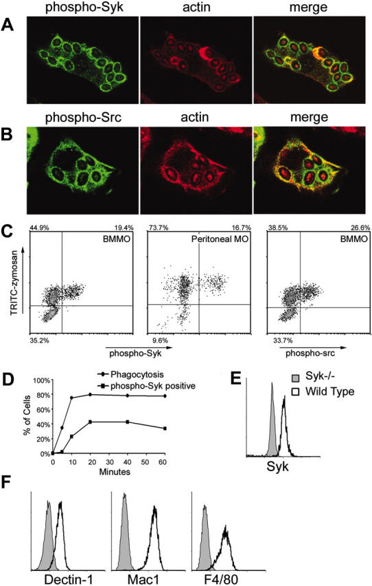
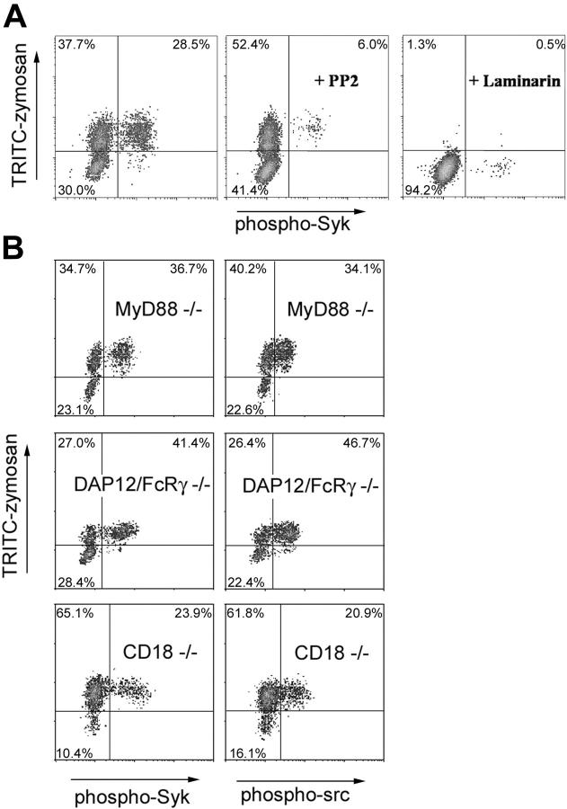
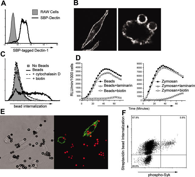
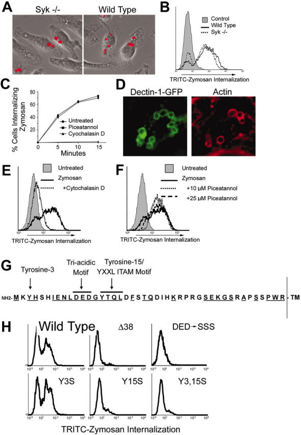
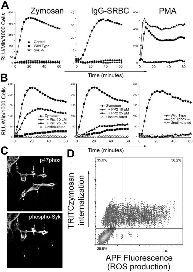
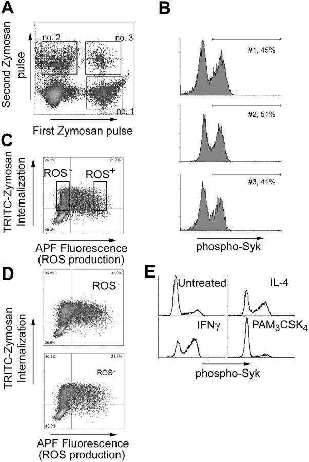
Dectin-1 is a lectin receptor for beta-glucan that is important for innate macrophage recognition of fungi and contributes to phagocytosis, reactive oxygen production, and induction of inflammatory cytokines. The mechanisms by which Dectin-1 mediates intracellular signaling are just beginning to be defined. Spleen tyrosine kinase (Syk) is a protein tyrosine kinase that is critical for adaptive immune responses where it mediates signaling through B-cell receptors, T-cell receptors, and Fc receptors. Here we report that Dectin-1 activates Syk in macrophages and is important for Dectin-1-stimulated reactive oxygen production, but not for phagocytosis. Syk activation is restricted to a subpopulation of macrophages that is in equilibrium with cells that cannot activate the pathway. The proportion of macrophages using this signaling pathway can be modulated by cytokine treatment. Thus, Dectin-1 signaling reveals dynamic macrophage heterogeneity in inflammatory activation potential.


Figures

Figure 1.
Figure 1.
Src/Syk activation by zymosan. IFN-γ-primed bone marrow-derived macrophages were fed zymosan for 15 minutes in the presence of sodium orthovanadate and activation of Syk (A) and Src family (B) kinases was visualized by immunofluorescence microscopy using anti-phospho-Syk (Tyr519/520) and anti-phospho-Src (Tyr 416) antibodies, respectively. (C) Tyrosine phosphorylation of Syk (left panels) and Src family kinases (right panel) was measured by flow cytometry of bone marrow-derived (BMMO) or peritoneal macrophages (MO) stimulated as described with fluorescently labeled zymosan for 20 minutes. (D) Tyrosine phosphorylation of Syk was measured in IFN-γ-primed bone marrow-derived macrophages stimulated for the indicated times with zymosan. Phagocytosis was synchronized by centrifugation of zymosan particles onto the macrophages at 4°C, and then warming the cells to 37°C. (E) Syk expression was detected by intracellular staining and flow cytometry in IFN-γ-primed wild-type and Syk-/- bone marrow-derived macrophages. (F) Surface expression of Dectin-1, Mac-1 (CD11b), and F4/80 on IFN-γ-primed bone marrow-derived macrophages was measured by flow cytometry.
Figure 2.
Figure 2.
Requirements for zymosan activation of Src/Syk signaling. (A) Syk activation was assessed in IFN-γ-primed bone marrow-derived macrophages stimulated with zymosan in the presence of the Src kinase inhibitor, PP2 (25 μM), or the soluble β-glucan, laminarin (0.5 mg/mL). (B) Syk and Src activation was measured in IFN-γ-primed bone marrow-derived macrophages obtained from mice deficient in MyD88, DAP12/FcRγ chain, or CD18 as indicated.
Figure 3.
Figure 3.
Dectin-1 signaling is sufficient to activate Syk in a subpopulation of macrophages. (A) Expression of SBP-tagged Dectin-1 in a stably transfected population of RAW264.7 macrophages was assessed by flow cytometry using an antibody to the epitope tag. (B) SBP-tagged Dectin-1 was localized by immunofluorescence microscopy in resting cells (left panel) or in cells fed streptavidin-coated beads (right panel). (C) RAW264.7 cells were incubated with streptavidin-coated beads in the presence of cytochalasin D (2.5 μM) or biotin (100 μM) as indicated, and internalization of the particles was measured by flow cytometry. (D) RAW264.7 cells expressing SBP-tagged Dectin-1 were incubated with streptavidin-coated beads (left panel) or zymosan (right panel) in the presence of laminarin (0.5 mg/mL) or biotin (100 μM) as indicated. ROS production was measured by luminol-enhanced chemiluminescence. (E) RAW264.7 cells expressing SBP-tagged Dectin-1 were stimulated with streptavidin-coated beads for 20 minutes in the presence of sodium orthovanadate, and activation of Syk was visualized by immunofluorescence microscopy as in Figure 1. (F) Activation of Syk in RAW264.7 cells expressing SBP-tagged Dectin-1 and stimulated with streptavidin-coated beads was assessed by flow cytometry.
Figure 4.
Figure 4.
Syk is not required for phagocytosis of zymosan. Wild-type and Syk-/- bone marrow-derived macrophages were cultured with fluorescently labeled zymosan for 15 minutes. Phagocytosis of zymosan was detected microscopically (A) and quantified by flow cytometry (B). (C) Bone marrow-derived macrophages were cultured with fluorescently labeled zymosan for the indicated times in the presence or absence of the Syk inhibitor piceatannol (25 μM) or the actin-disrupting agent cytochalasin D (2.5 μM). Phagocytosis was measured by flow cytometry, and the percentage of cells ingesting particles is shown. (D) HEK293 cells expressing GFP-tagged Dectin-1 were cultured with zymosan particles for 15 minutes, and Dectin-1 and actin were detected by immunofluorescence microscopy. (E) GFP/Dectin-expressing HEK293 cells were cultured with fluorescently labeled zymosan for 30 minutes in the presence or absence of cytochalasin D (E) or piceatannol (F), and internalization was measured by flow cytometry. (G) The cytoplasmic tail of murine Dectin-1 is depicted showing the 2 tyrosine residues and the triacidic motif. Residues that are conserved between mouse and human Dectin-1 are underlined. (H) HEK293 cells were transiently transfected with GFP-tagged Dectin-1 (wild type and the indicated mutants). The cells were cultured with fluorescently labeled zymosan, and internalization was measured by flow cytometry. The data are gated on GFP+ (Dectin-1-expressing) cells.
Figure 5.
Figure 5.
Syk is required for zymosan-induced ROS production. (A) IFN-γ-primed wild-type or Syk-/- bone marrow-derived macrophages were stimulated with zymosan, IgG-opsonized sheep red blood cells (SRBCs), or phorbol myristate acetate (PMA) as indicated, and generation of reactive oxygen was measured by enhanced chemiluminescence. (B) IFN-γ-primed wild-type or gp91phox-/- bone marrow-derived macrophages were stimulated with zymosan in the presence or absence of the Syk inhibitor, piceatannol (Pic), or the Src family kinase inhibitor, PP2, and generation of reactive oxygen was measured. (C) IFN-γ-primed bone marrow-derived macrophages were fed zymosan for 15 minutes, and translocation of p47phox to phagosome membranes and Syk phosphorylation was observed by immunofluorescence microscopy. (D) RAW264.7 macrophages expressing SBP-tagged Dectin-1 were stimulated with fluorescently labeled zymosan for 1 hour, and ROS production was detected by flow cytometry using APF.
Figure 6.
Figure 6.
The ability of Dectin-1 to activate Syk and ROS production is a transient property of a macrophage. (A) IFN-γ-primed bone marrow-derived macrophages were stimulated for 20 minutes with TRITC-labeled zymosan (first zymosan pulse), washed, and then stimulated with a second round of R-PE-labeled zymosan for an additional 207 minutes (second zymosan pulse). Populations of cells internalizing zymosan in the first, second, and both periods were identified by flow cytometry. (B) Syk activation was measured in cells using the gates indicated in panel A. (C) RAW264.7 cells expressing SBP-tagged Dectin-1 were stimulated with TRITC-zymosan for 1 hour, and ROS production was measured by flow cytometry usingAPF. Cells internalizing zymosan but not activating ROS production (ROS-) and cells in which ROS production was activated (ROS+) were sorted and expanded for 5 days in culture. (D) The sorted cells from panel C were restimulated with TRITC-zymosan and ROS production was again measured by flow cytometry. (E) Bone marrow-derived macrophages were treated overnight with IFN-γ (25 U/mL), IL-4 (20 ng/mL), or PAM3CSK4 (100 ng/mL) as indicated, and zymosan-induced activation of Syk was measured by flow cytometry. Data are gated on cells that had eaten zymosan.

References

    1. Janeway CA Jr, Medzhitov R. Innate immune recognition. Annu Rev Immunol. 2002;20: 197-216. - PubMed
    1. Takeda K, Akira S. Toll-like receptors in innate immunity. Int Immunol. 2005;17: 1-14. - PubMed
    1. Chu DH, Morita CT, Weiss A. The Syk family of protein tyrosine kinases in T-cell activation and development. Immunol Rev. 1998;165: 167-180. - PubMed
    1. Turner M, Schweighoffer E, Colucci F, Di Santo JP, Tybulewicz VL. Tyrosine kinase SYK: essential functions for immunoreceptor signaling. Immunol Today. 2000;21: 148-154. - PubMed
    1. Kiefer F, Brumell J, Al-Alawi N, et al. The Syk protein tyrosine kinase is essential for Fcgamma receptor signaling in macrophages and neutrophils. Mol Cell Biol. 1998;18: 4209-4220. - PMC - PubMed

Publication types

MeSH terms