

Case Reports
Intraparotid facial nerve schwannoma: a report of five cases and an analysis of MR imaging results
Affiliations
- PMID: 15956491
- PMCID: PMC8149055
Item in Clipboard
Case Reports
Intraparotid facial nerve schwannoma: a report of five cases and an analysis of MR imaging results
AJNR Am J Neuroradiol.
2005 Jun-Jul.
Abstract
We present five cases of intraparotid facial nerve schwannoma. In four of the five cases, tumors arising from the main facial nerve trunk were centered just below the skull base near the stylomastoid foramen and had a small cranial extension into the lower facial nerve canal. In three cases, each tumor had higher signal intensity around the periphery on T2-weighted images (target sign). These findings may be highly suggestive of an intraparotid facial nerve schwannoma.
Figures
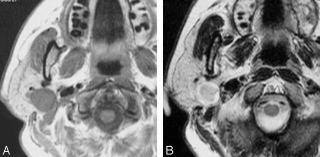
Case 1. A, Axial T1-weighted image (spin-echo: TR/TE, 650/10.7) shows a low-signal-intensity well-defined mass in the right parotid gland. B, Axial T2-weighted image (fast spin-echo: TR/TE, 3150/108) shows peripheral high signal intensity surrounding a central region of lower signal intensity.
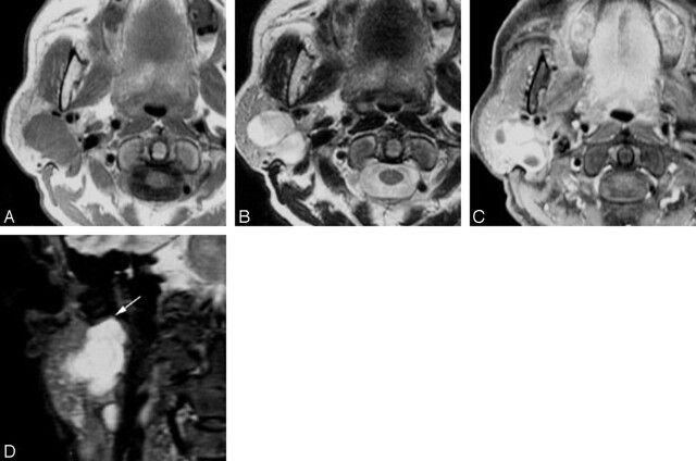
Case 2. A, Axial T1-weighted image (spin-echo: TR/TE, 650/9.3) shows a lobulated mass in the right parotid gland, with a low-signal-intensity well-defined mass in the right parotid gland. B, Axial T2-weighted image (fast spin-echo: TR/TE, 3150/108) shows a heterogeneous hyperintense mass. C, Axial gadolinium-enhanced T1-weighted image (spin echo; TR/TE, 650/9.3) shows a mass in the right parotid gland with heterogeneous enhancement. D, Coronal STIR image (TR/TE/TI, 3216/36/165) shows a hyperintense mass in the right parotid gland. The tumor is situated directly caudal to the stylomastoid foramen and protrudes into it (arrow).
References
-
- Balle VH, Greisen O. Neurilemomas of the facial nerve presenting as parotid tumors. Ann Otol Rhinol Laryngol 1984;93:70–72 - PubMed
-
- Forton GE, Moeneclaey LL, Offeciers FE. Facial nerve neuroma: report of two cases including histological and radiological imaging studies. Eur Arch Otorhinolaryngol 1994;251:17–22 - PubMed
-
- Chiang CW, Chang YL, Lou PJ. Multicentricity of intraparotid facial nerve schwannomas. Ann Otol Rhinol Laryngol 2001;110:871–874 - PubMed
-
- Chung SY, Kim DI, Lee BH, Yoon PH, Jeon P, Chung TS. Facial nerve schwannomas: CT and MR findings. Yonsei Med J 1998;39:148–153 - PubMed
-
- Martin N, Sterkers O, Mompoint D, Nahum H. Facial nerve neuromas: MR imaging—report of four cases. Neuroradiology 1992;34:62–67 - PubMed
Publication types
MeSH terms
LinkOut - more resources
Full Text Sources
Medical