

MR angiography at 3T versus digital subtraction angiography in the follow-up of intracranial aneurysms treated with detachable coils
- PMID: 15956496
- PMCID: PMC8149092
MR angiography at 3T versus digital subtraction angiography in the follow-up of intracranial aneurysms treated with detachable coils
Abstract
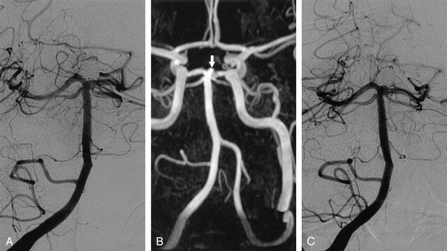
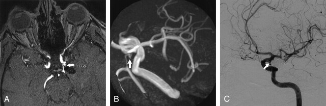
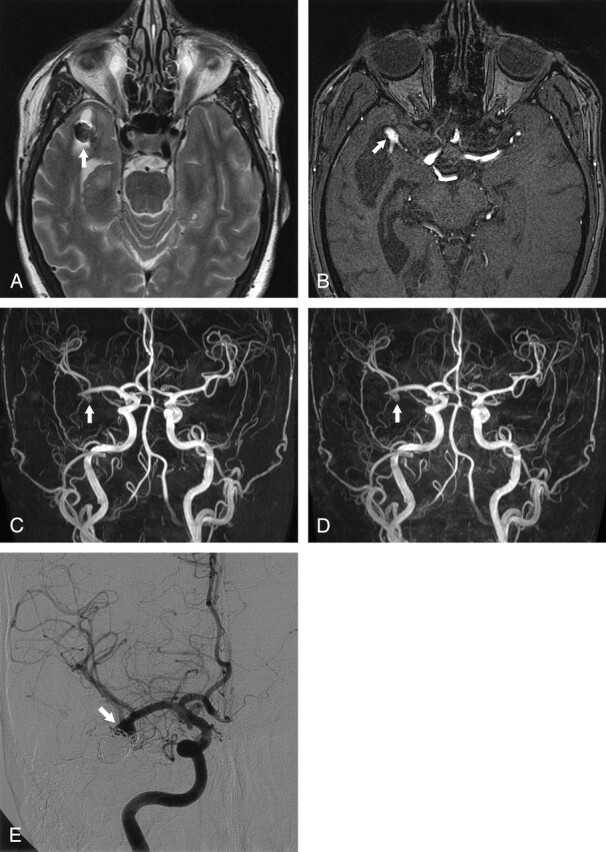
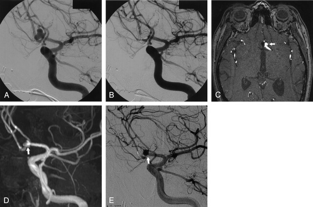
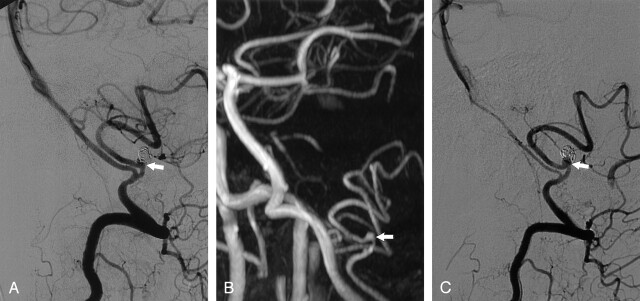
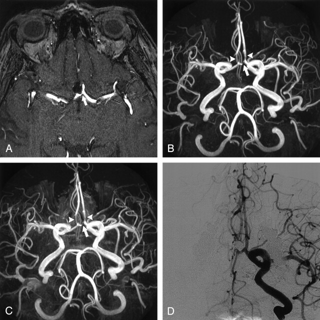
Background and purpose: Digital subtraction angiography (DSA) is used to follow-up intracranial aneurysms treated with detachable coils to identify recurrence and determine need for additional treatment. However, DSA is invasive and involves a small risk of neurologic complications. We assessed the feasibility and usefulness of 3D time-of-flight (TOF) MR angiography (MRA) performed at 3T compared with DSA for the follow-up of coil-treated intracranial aneurysms.
Methods: In a prospective study, 20 consecutive patients with 21 intracranial aneurysms treated with coils underwent DSA and nonenhanced and enhanced multiple overlapping thin-slab acquisition 3D TOF MRA at 3T on the same day at a mean follow-up of 6 months (range, 4-14 months) after coil placement. MRA images were evaluated for presence of artifacts, presence and size of aneurysm remnants and recurrences, patency of parent and branch vessels, and added value of contrast material enhancement. MRA and DSA findings were compared.
Results: Interobserver agreement of MRA was good, as was agreement between MRA and DSA. All three recurrences that needed additional treatment were detected with MRA. Minor disagreement occurred in four cases: three coil-treated aneurysms were scored on MRA images as having a small remnant, whereas on DSA images these aneurysms were occluded; the other aneurysm was scored on MRA images as having a small remnant, whereas on DSA images this was a small recurrence. Use of contrast material had no additional value. Coil-related MR imaging artifacts were minimal and did not interfere with evaluation of the occlusion status of the aneurysm.
Conclusion: High-spatial-resolution 3D TOF MRA at 3T is feasible and useful in the follow-up of patients with intracranial aneurysms treated with coil placement.
Figures
Similar articles
-
Time-of-flight MR angiography at 3T versus digital subtraction angiography in the imaging follow-up of 51 intracranial aneurysms treated with coils.Eur J Radiol. 2009 Dec;72(3):365-9. doi: 10.1016/j.ejrad.2008.08.005. Epub 2008 Sep 21. Eur J Radiol. 2009. PMID: 18809272 Clinical Trial.
-
Contrast-Enhanced and Time-of-Flight MRA at 3T Compared with DSA for the Follow-Up of Intracranial Aneurysms Treated with the WEB Device.AJNR Am J Neuroradiol. 2016 Sep;37(9):1684-9. doi: 10.3174/ajnr.A4791. Epub 2016 Apr 21. AJNR Am J Neuroradiol. 2016. PMID: 27102311 Free PMC article.
-
The effectiveness of 3T time-of-flight magnetic resonance angiography for follow-up evaluations after the stent-assisted coil embolization of cerebral aneurysms.Acta Radiol. 2014 Jun;55(5):604-13. doi: 10.1177/0284185113502335. Epub 2013 Sep 3. Acta Radiol. 2014. PMID: 24003259
-
MRA versus DSA for the follow-up imaging of intracranial aneurysms treated using endovascular techniques: a meta-analysis.J Neurointerv Surg. 2019 Oct;11(10):1009-1014. doi: 10.1136/neurintsurg-2019-014936. Epub 2019 May 2. J Neurointerv Surg. 2019. PMID: 31048457 Review.
-
MRA versus DSA for follow-up of coiled intracranial aneurysms: a meta-analysis.AJNR Am J Neuroradiol. 2014 Sep;35(9):1655-61. doi: 10.3174/ajnr.A3700. Epub 2013 Sep 5. AJNR Am J Neuroradiol. 2014. PMID: 24008171 Free PMC article.
Cited by
-
Outcomes of endovascular treatments of aneurysms: observer variability and implications for interpreting case series and planning randomized trials.AJNR Am J Neuroradiol. 2012 Apr;33(4):626-31. doi: 10.3174/ajnr.A2848. Epub 2011 Dec 22. AJNR Am J Neuroradiol. 2012. PMID: 22194386 Free PMC article.
-
The relation between packing and reopening in coiled intracranial aneurysms: a prospective study.Neuroradiology. 2005 Dec;47(12):942-5. doi: 10.1007/s00234-005-1446-9. Neuroradiology. 2005. PMID: 16136261 Clinical Trial.
-
CFD analysis in an anatomically realistic coronary artery model based on non-invasive 3D imaging: comparison of magnetic resonance imaging with computed tomography.Int J Cardiovasc Imaging. 2008 Apr;24(4):411-21. doi: 10.1007/s10554-007-9275-z. Epub 2007 Oct 23. Int J Cardiovasc Imaging. 2008. PMID: 17955344
-
Usefulness of Silent MR Angiography for Intracranial Aneurysms Treated with a Flow-Diverter Device.AJNR Am J Neuroradiol. 2019 May;40(5):808-814. doi: 10.3174/ajnr.A6047. Epub 2019 May 2. AJNR Am J Neuroradiol. 2019. PMID: 31048297 Free PMC article.
-
Aneurysm Recurrence Volumetry Is More Sensitive than Visual Evaluation of Aneurysm Recurrences.Clin Neuroradiol. 2016 Mar;26(1):57-64. doi: 10.1007/s00062-014-0330-6. Epub 2014 Aug 27. Clin Neuroradiol. 2016. PMID: 25159038 Clinical Trial.
References
-
- Molyneux A, Kerr R, Stratton I, et al, International Subarachnoid Aneurysm Trial (ISAT) Collaborative Group. International Subarachnoid Aneurysm Trial (ISAT) of neurosurgical clipping versus endovascular coiling in 2143 patients with ruptured intracranial aneurysms: a randomised trial. Lancet 2002;360:1267–1274 - PubMed
-
- Cognard C, Weill A, Spelle L, et al. Long-term angiographic follow-up of 169 intracranial aneurysms occluded with detachable coils. Radiology 1999;212:348–356 - PubMed
-
- Thornton J, Debrun GM, Aletich VA, Bashir Q, Charbel FT, Ausman JI. Follow-up angiography of intracranial aneurysms treated with endovascular placement of Guglielmi detachable coils. Neurosurgery 2002;50:239–249 - PubMed
-
- Sluzewski M, van Rooij WJ, Rinkel GJE, Wijnalda D. Endovascular treatment of ruptured intracranial aneurysms with detachable coils: long-term clinical and serial angiographic results. Radiology 2003;227:720–724 - PubMed
-
- Raymond J, Guilbert F, Weill A, et al. Long-term angiographic recurrences after selective endovascular treatment of aneurysms with detachable coils. Stroke 2003;34:1398–1403 - PubMed
Publication types
MeSH terms
LinkOut - more resources
Full Text Sources
Medical