. 2005 Jun-Jul;26(6):1399-404.

Endovascular treatment of high-flow carotid cavernous fistulas by stent-assisted coil placement

Affiliations

Endovascular treatment of high-flow carotid cavernous fistulas by stent-assisted coil placement

Fanny E Morón et al. AJNR Am J Neuroradiol. 2005 Jun-Jul.

Abstract

Background and purpose: Endovascular techniques are the methods of choice for the treatment of patients with carotid cavernous fistulas. We report our experience using stent-assisted coil placement for treatment of patients with high-flow fistulas that are associated with severe laceration of the internal carotid artery.

Methods: In a retrospective review of an internal endovascular therapy database covering the interval between October 2001 and October 2003, we identified a total of 5 patients presenting with 6 high-flow type A carotid cavernous fistulas (one had a bilateral fistula) that were associated with severe laceration of the internal carotid artery. All were treated first with stenting of the injured segment of the internal carotid artery followed by transarterial (3/6) and/or transvenous (4/6) obliteration of the fistula with detachable platinum coils. In 2 cases, a liquid adhesive was also used. In all instances, a compliant balloon was inflated within the stented arterial segment during coil deposition to avoid extension of coils into the parent artery.

Results: All 6 fistulas were obliterated, and each internal carotid artery was successfully reconstructed. Except for posttraumatic cranial nerve dysfunction in 1 patient, clinical outcome was very good. Follow-up angiograms in 3 of the 6 patients obtained at intervals between 3 and 6 months (mean, 4.5 months) revealed no fistula recurrence and no evidence of intimal hyperplasia within the stent.

Conclusion: In this series of patients with high-flow carotid cavernous fistula associated with severe injury to the internal carotid artery, stent-assisted coil placement offered a safe and effective treatment. Stent-assisted coil placement may increase the ability to successfully treat fistulas with severe injury to the internal carotid artery with preservation of the parent artery.


Figures

F<sc>ig</sc> 1.
Fig 1.
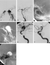
20-year-old woman (patient #3, Table 1) injured in an equestrian accident with multiple skull base fractures and bilateral carotid cavernous fistulas. A, Lateral projection of right common carotid artery arteriogram shows type A carotid cavernous fistula with no filling of the intradural segment of the internal carotid artery. Note prominent intracranial and orbital venous drainage. B, Lateral projection of right vertebral artery arteriogram demonstrates forward filling into right internal carotid artery. No demonstration of the communication between the proximal and distal internal carotid artery is seen. C, Lateral projection of right internal carotid artery angiogram following stent deployment with reconstruction of the vessel shows that there is now brisk antegrade filling of the internal carotid artery distal to the site of the fistula. D, Lateral projection of left internal carotid artery arteriogram shows type A carotid cavernous fistula. Although there is antegrade flow distal to the site of the fistula, the course and caliber of this segment of the internal carotid artery could not be determined. E, Lateral projection of left internal carotid artery arteriogram after stent deployment shows improved visualization of the cavernous segment of the internal carotid artery. Venous drainage to orbital veins persists. F, Lateral unsubtracted projection demonstrates bilateral stents in place (arrows). G, Lateral right internal carotid artery angiogram, unsubtracted, shows an inflated balloon inside the stent (arrow). This was used during coil deposition. H, Lateral projection of left internal carotid artery arteriogram demonstrates persistent flow after extensive coiling. I, Lateral projection of left internal carotid artery arteriogram shows a small aliquot of liquid adhesive (arrow) that was instilled in the proximal portion of the superior ophthalmic vein. This resulted in complete closing of the fistula. J-K, Frontal and lateral projections of right internal carotid artery (J) and left internal carotid artery (K) on 3-month follow-up arteriogram demonstrate patency of both internal carotid arteries with antegrade filling of their branches, dense coil packing, and complete closure of the fistulas.
F<sc>ig</sc> 2.
Fig 2.
62-year-old woman (patient 5, Table 1) with sudden left-sided ptosis. A, Frontal projection of left internal carotid artery arteriogram confirms type A carotid cavernous fistula with poor antegrade filling of the internal carotid artery branches. Note extensive venous drainage to superior ophthalmic vein, pterygopalatine plexus, and both inferior petrosal sinus and contralateral cavernous sinus. B, Lateral projection of left vertebral artery arteriogram does not demonstrate communication between the proximal and distal internal carotid artery segments. The right internal carotid artery arteriogram (not shown) also did not demonstrate a connection between these two segments. C, Lateral unsubtracted projection demonstrates the stent in place (arrow). D, Lateral road map image of left internal carotid artery arteriogram shows inflated balloon in stent (triple black arrows), used during coiling to prevent coils from coming into the arterial lumen, which could not be visualized. Note microcatheter in inferior petrosal sinus (white arrow) for transvenous embolization. E, Lateral projection of left internal carotid artery arteriogram after extensive coiling with almost complete obliteration of the aneurysm and carotid cavernous fistula. Note small residual aneurysm (arrow) and antegrade filling of the internal carotid artery branches. No additional coils could be placed. F, Lateral projection of left internal carotid artery on 6-month follow-up arteriogram demonstrates stable appearance of the aneurysm and carotid cavernous fistula with normal antegrade filling of the internal carotid artery. G, In lateral unsubtracted projection of left internal carotid artery on 6-month follow-up arteriogram, the stent demonstrates the lumen (arrow) of the internal carotid artery.

References

    1. Barrow DL, Spector RH, Braun IF, Tindall SC, Tindall GT. Classification and treatment of spontaneous carotid-cavernous sinus fistulas. J Neurosurg 1985;62:248–56 - PubMed
    1. Debrun GM, Vinuela F, Fox AJ, Drake KR, Ahn HS. Indications for treatment and classification of 132 carotid-cavernous fistulas. Neurosurgery 1988;22:285–289 - PubMed
    1. Lewis A, Tomsick TA, Tew JJ. Management of 100 consecutive direct carotid cavernous fistulas: results of treatment with detachable balloons. Neurosurgery 1995;36:239–244 - PubMed
    1. Lee CY, Yim MB, Kim IM, Son EI, Kim DW. Traumatic aneurysm of the supraclinoid internal carotid artery and an associated carotid-cavernous fistula: vascular reconstruction performed using intravascular implantation of stents and coils—case report. J Neurosurg 2004;100:115–119 - PubMed
    1. Ahn JY, Lee BH, Joo JY. Stent-assisted Guglielmi detachable coils embolization for the treatment of a traumatic carotid cavernous fistula. J Clin Neurosci 2003;10:96–98 - PubMed

LinkOut - more resources