Stereolithographic vascular replicas from CT scans: choosing treatment strategies, teaching, and research from live patient scan data
- PMID: 15956511
- PMCID: PMC8149057
Stereolithographic vascular replicas from CT scans: choosing treatment strategies, teaching, and research from live patient scan data
Abstract
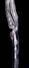
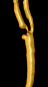
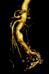
Our goal was to develop a system that would allow us to recreate live patient arterial pathology by using an industrial technique known as stereolithography (or rapid prototyping). In industry, drawings rendered into dicom files can be exported to a computer programmed to drive various industrial tools. Those tools then make a 3D structure shown by the original drawings. We manipulated CT scan dicom files to drive a stereolithography machine and were able to make replicas of the vascular diseases of three patients.
Figures


References
-
- Webb PA. A review of rapid prototyping (RP) techniques in the medical and biomedical sector. J Med Eng Technol 2000;24:149–153 - PubMed
-
- Brown GA, Firoozbakhsh K, DeCoster TA, et al. Rapid prototyping: the future of trauma surgery? J Bone Joint Surg Am 2003;85-A (suppl 4):49–55 - PubMed
-
- Potamianos P, Amis AA, Forester AJ, et al. Rapid prototyping for orthopaedic surgery. Proc Inst Mech Eng [H]. 1998;212:383–393 - PubMed
-
- Girod S, Teschner M, Schrell U, et al. Computer-aided 3-D simulation and prediction of craniofacial surgery: a new approach. J Craniomaxillofac Surg 2001;29:156–158 - PubMed
-
- Petzold R, Zeilhofer H, Kalender WA. Rapid protyping technology in medicine: basics and applications. Comput Med Imaging Graph 1999;23:277–284 - PubMed
MeSH terms
LinkOut - more resources
Full Text Sources
Other Literature Sources
Medical
