. 2005 Jun-Jul;26(6):1551-62.

Diffusion-weighted and fluid-attenuated inversion recovery imaging in Creutzfeldt-Jakob disease: high sensitivity and specificity for diagnosis

Affiliations

Diffusion-weighted and fluid-attenuated inversion recovery imaging in Creutzfeldt-Jakob disease: high sensitivity and specificity for diagnosis

Geoffrey S Young et al. AJNR Am J Neuroradiol. 2005 Jun-Jul.

Abstract

Background and purpose: Abnormalities on diffusion-weighted images (DWIs) and fluid-attenuated inversion recovery (FLAIR) images are reported in Creutzfeldt-Jakob disease (CJD). To our knowledge, no large study has been conducted to determine the sensitivity and specificity of DWI and FLAIR imaging for diagnosing CJD.

Methods: Two neuroradiologists, blinded to diagnosis, retrospectively evaluated DWI and FLAIR images from 40 patients with probable or definite CJD and 53 control subjects with other forms of dementia and rated the likelihood of CJD on the basis of the imaging findings.

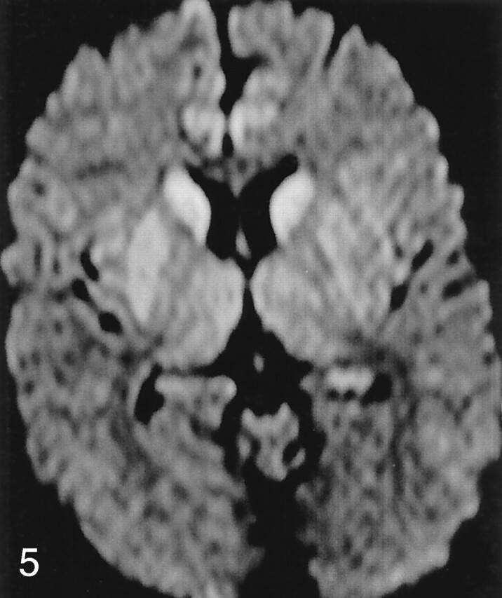
Results: DWI and FLAIR imaging was 91% sensitive, 95% specific, and 94% accurate for CJD. Interrater reliability was high (kappa = 0.93). Sensitivity was higher for DWI than FLAIR imaging. Abnormalities involved cortex and deep gray matter (striatum and/or thalamus) in 68% of patients with CJD, cortex alone in 24%, and deep gray matter alone in 5%. The most typical and specific patterns were corresponding hyperintensity on both FLAIR images and DWIs confined to the gray matter in the cortex, striatum, medial and/or posterior thalamus, or a combination of these areas. Narrow-window soft-copy review of artifact-free DWIs and FLAIR images and recognition of the normal variation in cortical signal intensity proved critical for successful differentiation of CJD from other dementias.

Conclusion: Because specific patterns of abnormality on DWI and FLAIR images are highly sensitive and specific for CJD, these sequences should be performed whenever CJD is suspected.


Figures

F<sc>ig</sc> 1.
Fig 1.
Normal variations, which can complicate assessment of pathologic cortical hyperintensity on FLAIR and DWI. A, Axial FLAIR (TR/TE/TI = 10000/140/2200) image shows insular cortex slightly hyperintense (arrows) to neocortex. B, More superior axial FLAIR image shows relative hyperintensity in cingulate cortex (arrows). C, Axial DWI (TR/TE = 8000/minimal, b = 1000 s/mm2, same level as A) shows hyperintensity in bilateral insular cortex (arrows). D, More superior DWI (same level as B) shows relative hyperintensity cingulate cortex (arrows), accentuated by frontal magnetic susceptibility artifact.
F<sc>ig</sc> 2.
Fig 2.
A 50-year-old man with definite sCJD. A, Axial DWI shows pathologic hyperintensity in bilateral posterior temporoparietal neocortex. Cortex along parieto-occipital fissure is abnormally hyperintense (vertical arrows), but primary visual region is spared (horizontal arrows). Note asymmetric abnormal hyperintensity in right cingulum (arrowhead). Striatum is uninvolved. B, FLAIR image at same level shows more subtle pathologic hyperintensity in all abnormal regions on DWI, as shown in cingulate cortex (arrowhead).
F<sc>ig</sc> 3.
Fig 3.
A 52-year-old man with probable sCJD. A, Axial DWI shows pathologic hyperintensity in bilateral parietal neocortex and sparing of cortex in postcentral gyrus along the central sulcus (arrows) and entire precentral gyrus. Note subtle abnormality of paramedian frontal cortex (arrowheads). B, More superior DWI shows relatively sparing of cortex on both sides of central sulcus (arrows). C, More superior DWI confirms identification of central sulcus (arrows) and sparing of precentral and postcentral gyri.
F<sc>ig</sc> 4.
Fig 4.
A 26-year-old man with sCJD. A, Axial DWI shows extensive, asymmetric, right-greater-than-left neocortical involvement and abnormal hyperintensity of right caudate nucleus, putamen, and thalamus (arrowheads). Left caudate nucleus may be mildly hyperintense, but left putamen and thalamus are not definitely abnormal. B, More superior axial DWI shows involvement of posterior frontal (top arrowhead) and anterior parietal (bottom arrowhead) cortex, with sparing of cortex on edges of the central sulcus (arrow).
F<sc>ig</sc> 5.
Fig 5.
Striatal without neocortical involvement. Axial DWI in a 40-year-old man with sCJD shows abnormal symmetric hyperintensity in bilateral caudate nuclei, right putamen, and possibly right thalamus. Left putamen and thalamus are not definitely abnormal. Mild hyperintensity in bilateral cingulate cortex is thought to be normal variation and magnetic susceptibility artifact.
F<sc>ig</sc> 6.
Fig 6.
Axial DWI in a 78-year-old woman with sCJD shows symmetric hyperintensity in bilateral caudate nuclei and anterior putamina. No definite neocortical involvement is seen, as temporal hyperintensity (arrowheads) is thought to be magnetic susceptibility artifact, and appearance of insular and cingulate cortices may be within normal limits.
F<sc>ig</sc> 7.
Fig 7.
Abnormal thalamic appearance in sCJD. A, DWI in a 41-year-old man shows symmetric hyperintensity in bilateral caudate nuclei and putamina and medial and posterior thalami (double hockey stick). No definite neocortical abnormality is seen. Cingulate cortex and insular cortices are mildly hyperintense but likely within normal limits. B, Axial DWI in a 59-year-old woman shows abnormal hyperintensity in bilateral medial thalami and pulvinar. Left insular cortex (arrows) was thought to be definitely abnormal, and right, possibly abnormal. Caudate nuclei and putamina are spared.
F<sc>ig</sc> 8.
Fig 8.
False-positive CJD. Patient had mild cognitive impairment. A, On DWI, signal intensities of insular cortices (arrows) and dorsomedial thalami (arrowheads) were called abnormal. B, More superior DWI shows no clear abnormality, although subtle hyperintensity in caudate nuclei is questioned. Magnetic susceptibility artifact somewhat obscure frontal lobes. C, More superior DWI suggests abnormal hyperintensity of medial frontal lobes.
F<sc>ig</sc> 9.
Fig 9.
False-negative CJD. A, Axial DWI (poor windows, film cohort) in a 65-year-old man with definite sCJD retrospectively shows subtle hyperintensity of bilateral caudate heads and anterior putamina. Medial and dorsal thalami are subtly abnormal but more diffusely than is typical of CJD. Insular cortex may be pathologically hyperintense or normal. B, Even in retrospect, calling an abnormality on corresponding FLAIR image (mildly motion degraded) is difficult.
F<sc>ig</sc> 10.
Fig 10.
False-negative CJD (film cohort). A, Axial DWI in a 49-year-old man with definite sCJD shows no clear abnormality. Image is motion degraded and poorly windowed, with severe magnetic susceptibility artifact in bifrontal regions. B, Corresponding FLAIR image shows marked prominence of ventricles and sulci for the patient’s age. In retrospect, extensive cortical thinning and probably pathologic hyperintensity are found. CJD not called because of lack of DWI confirmation and difficulty in assessing thin cortex in this patient (with clinical signs for >6 months).

References

    1. Prusiner SB. Prions. Proc Natl Acad Sci U S A 95:1998;13363–13383 - PMC - PubMed
    1. Geschwind MD, Jay C. Alzheimer’s Disease and Other Primary Dementias. In: Harrison’s Principles of Internal Medicine. Bird T, ed. McGraw Hill,2003. :2391–2398
    1. World Health Organization. Global surveillance, diagnosis, and therapy of human transmissible spongiform encephalopathies: report of a WHO consultation. In: Global Surveillance, Diagnosis, and Therapy of Human Transmissible Spongiform Encephalopathies. Geneva: World Health Organization;1998. :1–29
    1. Tschampa H. Patients with Alzheimer’s disease and dementia with Lewy bodies mistaken for Creutzfeldt-Jakob Disease. J Neurol Neurosurg Psychiatry 2001;71:33–39 - PMC - PubMed
    1. Zerr I, Pocchiari M, Collins S, et al. Analysis of EEG and CSF 14-3-3 proteins as aids to the diagnosis of Creutzfeldt-Jakob disease. Neurology 2000;55:811–815 - PubMed

Publication types