

Case Reports
Diffusion tensor imaging in lissencephaly
Affiliations
- PMID: 15956534
- PMCID: PMC8149082
Item in Clipboard
Case Reports
Diffusion tensor imaging in lissencephaly
AJNR Am J Neuroradiol.
2005 Jun-Jul.
Abstract
Lissencephaly is a rare brain malformation characterized histologically by arrested neuronal migration such that the brain resembles that of a fetus before 23-24 weeks gestation. We studied a neonate with lissencephaly by using diffusion tensor imaging and suggest the dysplastic densely cellular layer IV is visible as a band of anisotropic diffusion. Fiber tracking showed lack of connectivity between the cortex and deep white matter and an abnormal limbic system.
Figures
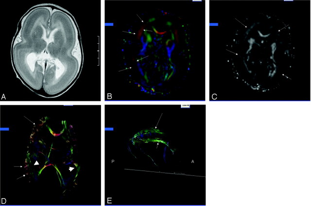
Patient, a 30-day-old male with isolated lissencephaly. A, Axial T2-weighted image shows diffuse thickening of the cortex with lack of sulcation. B, Axial 2D color map through the body of the lateral ventricles shows bands of color (arrows) corresponding to the deep cortex. C, Axial anisotropy map shows bands of higher anisotropy corresponding to the bands of color (arrows) seen on the color maps. D, Axial 3D presentation of corpus callosum, fronto-occipital fasciculus (short arrows), and presumed cell layer IV (arrows). E, Sagittal 3D depiction of the cingulum (long arrow) and fornix (short arrow). Note absence of the temporal projections of the cingulum and fornix.
References
-
- Dong Q, Welsh RC, Chenevert TL, et al. Clinical applications of diffusion tensor imaging. J Magn Reson Imaging 2004;19:6–18 - PubMed
-
- Ozarslan E, Mareci TH. Generalized diffusion tensor imaging and analytical relations between diffusion tensor imaging and high angular resolution imaging. Mag Reson Med 2003;50:955–965 - PubMed
-
- DaSilva AFM, Tuch DS, Weigell MR, Hadjikhani N. A primer on diffusion tenor imaging of anatomical substructures. Neurosurg Focus 2003;15:1–4 - PubMed
-
- Mori S, Crain BJ, Chacko VP, van Zijl. Three-dimensional tracking of axonal projections in the brain by magnetic resonance imaging. Ann Neurol 1999;45265–269 - PubMed
Publication types
MeSH terms
LinkOut - more resources
Full Text Sources
Medical