. 2005 Jul;79(13):8065-78.
doi: 10.1128/JVI.79.13.8065-8078.2005.

Gene 5 of the avian coronavirus infectious bronchitis virus is not essential for replication

Affiliations

Gene 5 of the avian coronavirus infectious bronchitis virus is not essential for replication

Rosa Casais et al. J Virol. 2005 Jul.

Abstract

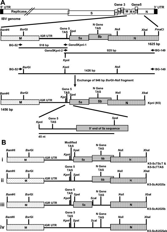
The avian coronavirus Infectious bronchitis virus (IBV), like other coronaviruses, expresses several small nonstructural (ns) proteins in addition to those from gene 1 (replicase) and the structural proteins. These coronavirus ns genes differ both in number and in amino acid similarity between the coronavirus groups but show some concordance within a group or subgroup. The functions and requirements of the small ns gene products remain to be elucidated. With the advent of reverse genetics for coronaviruses, the first steps in elucidating their role can be investigated. We have used our reverse genetics system for IBV (R. Casais, V. Thiel, S. G. Siddell, D. Cavanagh, and P. Britton, J. Virol. 75:12359-12369, 2001) to investigate the requirement of IBV gene 5 for replication in vivo, in ovo, and ex vivo. We produced a series of recombinant viruses, with an isogenic background, in which complete expression of gene 5 products was prevented by the inactivation of gene 5 following scrambling of the transcription-associated sequence, thereby preventing the expression of IBV subgenomic mRNA 5, or scrambling either separately or together of the translation initiation codons for the two gene 5 products. As all of the recombinant viruses replicated very similarly to the wild-type virus, Beau-R, we conclude that the IBV gene 5 products are not essential for IBV replication per se and that they are accessory proteins.


Figures

FIG. 1.
FIG. 1.
Schematic diagrams for the construction of the modified IBV gene 5 cDNAs. (A) A KpnI restriction endonuclease site was introduced by changing nucleotides 25454GTT to ACC proximal to the IBV Beaudette gene 5 TAS using overlapping PCR mutagenesis. This cDNA (K5) was used for introducing modifications into the gene 5 TAS (ScTScT and ScTTAS) or to scramble the ORF 5a ATG (ScAUG5a) by replacing the 45-nt KpnI-SpeI fragment with adapters containing the required nucleotide substitutions. The ORF 5b ATG was scrambled, and a ScaI restriction endonuclease site was introduced using overlapping PCR mutagenesis. The modified cDNA with the initiation codons of both ORF 5a and ORF 5b scrambled, ScAUG5ab, was constructed by replacing the SpeI-NsiI of ScAUG5a with the corresponding fragment from ScAUG5b. The overlapping PCR mutagenesis step for introduction of the KpnI restriction endonuclease site, in which the Beaudette 946-bp BsrGI-NsiI cDNA fragment was replaced with the corresponding PCR fragment containing the KpnI site, is shown. (B) Diagrams of the modified cDNAs that together with the K5 cDNA were used to produce the rIBVs. The positions of the IBV gene products M, 5a, 5b, and N are shown as labeled bars. However, the coding sequences for ORFs 5a and 5b are shown as black lines, following scrambling of the initiation codon, to indicate that the sequences are retained but that translation of the gene product is lost. IGR UTR, intergenic untranslated region.
FIG. 2.
FIG. 2.
Schematic diagrams showing the transient dominant selection process for modifying the full-length cDNA within the genome of vaccinia virus vNotI/IBVFL. The modified cDNAs shown in Fig. 1B were introduced as a 1,456-bp BamHI-XbaI fragment into the TDS GPT transfer/recombination vector pGPTNEB193rev. (A) Representation of one of the modified cDNAs, K5-ScAUG5b, in pGPTNEB193rev. (B) Outline of the two-step process for modifying the IBV cDNA within vNotI/IBVFL. In step 1, the complete plasmid DNA is integrated into the full-length IBV cDNA by a single-step homologous recombination event; a potential recombination event is shown. The resultant rVV has a GPT+ phenotype, allowing selection in the presence of mycophenolic acid. Removal of mycophenolic acid results in two types of spontaneous intramolecular recombination events, I and II, due to the instability of the IBV cDNA with tandem repeats of similar sequences; I results in reversion to the Beaudette genotype (no introduced modifications), and II results in introduction of the modification(s). Both recombination events result in the loss of GPT. The example shown outlines the introduction of the scrambled 5b AUG and associated nucleotide substitutions.
FIG. 3.
FIG. 3.
Northern blot analysis of IBV sg mRNAs 4 to 6 following infection of CK cells with the rIBVs. CK cells were infected with 3 × 10⁶ PFU of Beau-R or of each rIBV, and total cellular RNA was extracted at 24 h postinfection, separated by electrophoresis in 1% denaturing formaldehyde-agarose gels, and transferred to nylon membranes. The IBV-derived RNAs were detected nonisotopically with a mixture of two IBV-specific probes, a 666-bp IBV 3′ UTR probe and a 430-bp IBV N gene probe. RNAs from mock-infected CK cells (lane 1) or CK cells infected with Beau-R (lane 2), BeauR-K5-1 and -2 (lanes 3 and 4), BeauR-K5-ScTScT-1 and -2 (lanes 5 and 6), BeauR-K5-ScTTAS-1 and -2 (lanes 7 and 8), BeauR-K5-ScAUG5a-1 and -2 (lanes 9 and 10), BeauR-K5-ScAUG5b-1 and -2 (lanes 11 and 12), or BeauR-K5-ScAUG5ab-1 and -2 (lanes 13 and 14) were analyzed. The RNA species detected below sg mRNA 6 are observed routinely for all strains of IBV, as originally identified (50), and are of unknown origin.
FIG. 4.
FIG. 4.
Western blot analysis for the detection of IBV 5b following infection of CK cells with the rIBVs. CK cells were infected with Beau-R or with the two isolates of each type of rIBV. Proteins derived from the infected cell lysates were separated on Tris-Tricine 15% SDS-polyacrylamide gels, transferred to polyvinylidene difluoride membranes, and analyzed for the presence of IBV M and 5b proteins by ECL. The M protein was detected using a rabbit anti-M serum, and 5b was detected using rat anti-serum SK21 followed by goat anti-rabbit and goat anti-rat immunoglobulins conjugated to horseradish peroxidase. Panel A shows the analysis of cell lysates from uninfected CK cells (lane 1) or CK cells infected with Beau-R (lane 2), BeauR-K5-1 and -2 (lanes 3 and 4), BeauR-K5-ScTScT-1 and -2 (lanes 5 and 6), and BeauR-K5-ScTTAS-1 and -2 (lanes 7 and 8). Panel B shows the analysis of cell lysates from uninfected CK cells (lane 1) or CK cells infected with Beau-R (lane 2), BeauR-K5-ScAUG5a-1 and -2 (lanes 3 and 4), BeauR-K5-ScAUG5b-1 and -2 (lanes 5 and 6), or BeauR-K5-ScAUG5ab-1 and -2 (lanes 7 and 8). The proteins representing the IBV M protein (34 kDa) and the 5b product (9.7 kDa) are shown. Molecular weights were determined using broad-range SDS-polyacrylamide gel electrophoresis standard proteins (Bio-Rad).
FIG. 5.
FIG. 5.
Detection of 5b in infected Vero cells by indirect immunofluorescence. Vero cells at 70% confluency were infected with Beau-R or with one isolate of each type of rIBV and fixed 20 h postinfection using 4% paraformaldehyde. The infected cells were analyzed by indirect immunofluorescence using either rat SK21 5b anti-peptide sera raised against two KLH-conjugated peptides derived from 5b or mouse monoclonal antibody IE7 against 3c (E) followed by either Alexa 488-labeled donkey anti-rat antibody or Alexa 568-labeled goat anti-mouse antibody, respectively, and stained with ToPro 3 to visualize nuclear DNA. Panel A shows Vero cells that had been infected with Beau-R and analyzed using preimmune rat SK21 serum and anti-E monoclonal antibody IE7. Panel B shows mock-infected Vero cells analyzed with rat SK21 anti-peptide sera and anti-E monoclonal antibody IE7. The other panels show Vero cells infected with Beau-R (C and D), BeauR-K5-1 (E and F), BeauR-K5-ScTTAS-1 (G and H), BeauR-K5-ScTScT-1 (I and J), BeauR-K5-ScAUG5a-1 (K and L), BeauR-K5-ScAUG5b-1 (M and N), or BeauR-K5-ScAUG5ab-1 (O and P) analyzed with rat SK21 5b anti-peptide sera (C, E, G, I, K, M, and O) or mouse monoclonal antibody IE7 against 3c (D, F, H, J, L, N, and P). The cells were examined using confocal microscopy at ×40 magnification; red corresponds to the presence of IBV E protein and green to the presence of 5b protein.
FIG. 6.
FIG. 6.
Comparison of the in vitro growth kinetics of the gene 5 rIBVs in CK cells. Two independent rIBVs with each modification were used. CK cells were infected with 5.4 × 10⁴ PFU (MOI ≈ 0.03) of Beau-R or of each rIBV, and the titer of progeny virus was analyzed by plaque titration assay on CK cells over a period of 48 h. Panels show a comparison of the growth kinetics of each analogous pair (1 [dashed lines with open square] and 2 [dashed lines with triangle]) of the rIBVs with Beau-R (solid line and filled diamond). (A) BeauR-K5, (B) BeauR-K5-ScTTAS, (C) BeauR-K5-ScTScT, (D) BeauR-K5-ScAUG5a, (E) BeauR-K5-ScAUG5b, and (F) BeauR-K5-ScAUG5ab. The error bars represent standard deviations.
FIG. 7.
FIG. 7.
Comparison of the in ovo growth kinetics of the gene 5 rIBVs in 11-day-old embryonated eggs. Two independent rIBVs with each modification were used. The embryonated eggs were inoculated with 0.4 PFU of Beau-R or of each rIBV and moved to 4°C at different time points up to 40 h postinfection. Allantoic fluid from the eggs was analyzed for progeny virus by plaque titration assay on CK cells. The panels show the comparisons of the growth kinetics of each analogous pair(1 and 2 [dashed line and filled squares]) (except for BeauR-K5-ScAUG5b, in which only one virus was analyzed) of the rIBVs with Beau-R (solid line and filled diamonds). (A) BeauR-K5, (B) BeauR-K5-ScTScT, (C) BeauR-K5-ScTTAS, (D) BeauR-K5-ScAUG5a, (E) BeauR-K5-ScAUG5b, and (F) BeauR-K5-ScAUG5ab. The error bars represent standard deviations.
FIG. 8.
FIG. 8.
Ex vivo growth kinetics of the BeauR-K5-ScTScT gene 5 rIBVs in chicken tracheal organ cultures. Groups of five chicken TOCs were infected for 1 h at 37°C with 2.7 × 10⁴ PFU of Beau-R, rIBV BeauR-K5-1, and the two gene 5 rIBVs, BeauR-K5-ScTScT-1 and BeauR-K5-ScTScT-2, that are unable to generate the gene 5 sg mRNA 5. The medium was removed, the TOCs were washed three times with PBS, and incubation continued with 1 ml of medium. At selected time points, progeny viruses from three groups of TOCs were assayed by plaque titration on CK cells. The growth profiles of the four viruses are shown. The error bars represent standard deviations.

References

    1. Almazán, F., J. M. González, Z. Pénzes, A. Izeta, E. Calvo, J. Plana-Durán, and L. Enjuanes. 2000. Engineering the largest RNA virus genome as an infectious bacterial artificial chromosome. Proc. Natl. Acad. Sci. USA 97:5516-5521. - PMC - PubMed
    1. Ausubel, F. M., R. Brent, R. E. Kingston, D. D. Moore, J. G. Seidman, J. A. Smith, and K. Struhl. 1987. Current protocols in molecular biology. John Wiley and Sons, Inc., New York, N.Y.
    1. Bonfield, J. K., K. F. Smith, and R. Staden. 1995. A new DNA sequence assembly program. Nucleic Acids Res. 23:4992-4999. - PMC - PubMed
    1. Boulanger, D., P. Green, T. Smith, C.-P. Czerny, and M. A. Skinner. 1998. The 131-amino-acid repeat region of the essential 39-kilodalton core protein of fowlpox virus FP9, equivalent to vaccinia virus A4L protein, is nonessential and highly immunogenic. J. Virol. 72:170-179. - PMC - PubMed
    1. Britton, P., S. Evans, B. Dove, M. Davies, R. Casais, and D. Cavanagh. 2005. Generation of a recombinant avian coronavirus infectious bronchitis virus using transient dominant selection. J. Virol. Methods 123:203-211. - PMC - PubMed

Publication types