. 2005 Jul;79(13):8201-7.
doi: 10.1128/JVI.79.13.8201-8207.2005.

Differential sensitivity of murine leukemia virus to APOBEC3-mediated inhibition is governed by virion exclusion

Affiliations

Differential sensitivity of murine leukemia virus to APOBEC3-mediated inhibition is governed by virion exclusion

Brian P Doehle et al. J Virol. 2005 Jul.

Abstract

While members of the APOBEC3 family of human intrinsic resistance factors are able to restrict the replication of Vif-deficient forms of human immunodeficiency virus type 1 (HIV-1), they are unable to block replication of wild-type HIV-1 due to the action of Vif, which induces their degradation. In contrast, HIV-1 Vif is unable to block inhibition mediated by APOBEC3 proteins expressed by several heterologous species, including mice. Here, we have asked whether the simple retrovirus murine leukemia virus (MLV) is sensitive to restriction by the cognate murine or heterologous, human APOBEC3 proteins. We demonstrate that MLV is highly sensitive to inhibition by human APOBEC3G and APOBEC3B but resistant to inhibition by murine APOBEC3 or by other human APOBEC3 proteins, including APOBEC3F. This sensitivity fully correlates with the ability of these proteins to be packaged into MLV virion particles: i.e., human APOBEC3G and APOBEC3B are packaged while murine APOBEC3 and human APOBEC3F are excluded. Moreover, this packaging in turn correlates with the differential ability of these APOBEC3 proteins to bind MLV Gag. Together, these data suggest that MLV Gag has evolved to avoid binding, and hence virion packaging, of the cognate murine APOBEC3 protein but that MLV infectivity is still restricted by certain heterologous APOBEC3 proteins that retain this ability. Moreover, these results suggest that APOBEC3 proteins may help prevent the zoonotic infection of humans by simple retroviruses and provide a mechanism for how simple retroviruses can avoid inhibition by APOBEC3 family members.


Figures

FIG. 1.
FIG. 1.
Expression of mA3 mRNA in a variety of cell lines and murine tissues. (A) RT-PCR analysis using total RNA preparations from a variety of mouse tissues and cell lines. BW5147 is a murine T-cell lymphoma cell line, while 293T cells are of human origin and function as a negative control. Neg, no cDNA added; M, marker. (B) This RT-PCR amplification of mA3 mRNA was performed in parallel using increasing amounts of BW5147 cDNA under the same conditions as in panel A.
FIG. 2.
FIG. 2.
Comparison of the abilities of hA3G and mA3 to inhibit Vif-deficient HIV-1 or wild-type MLV infectivity. (A) 293T cells were transfected with the full-length MLV proviral clone pNCS together with the MLV-based luciferase expression vector pFB-Luc. Increasing levels of plasmids encoding mA3 or hA3G were cotransfected, with the parental pcDNA3 plasmid acting as a control (None) and used to maintain a constant level of DNA. Supernatant virions were collected and used to infect naïve 293T cells at 44 h after transfection. Induced luciferase levels were analyzed 24 h after infection. Data are presented as a percentage of the level of luciferase activity detected in cells infected with virions derived from cells that did not express an exogenous APOBEC3 protein. The average of three experiments with standard deviation is indicated. (B) Same as panel A, except that the 293T cells were transfected with the Vif-deficient HIV-1 luciferase indicator proviral clone pNL-HXB-LUCΔVIF in place of pNCS and pFB-Luc. (C) Western blot of the virus-producing 293T cells analyzed in panel A using antisera specific for the HA epitope tag present on the APOBEC3 proteins (αHA) or using an anti-MLV capsid antiserum (αCA).
FIG. 3.
FIG. 3.
hA3B, but not hA3F, inhibits MLV infectivity. One hundred twenty-five nanograms of each of the indicated APOBEC3 expression plasmids, or the pcDNA3 parental plasmid (None), was cotransfected into 293T cells along with pNCS and pFB-Luc, and the effect on MLV infectivity was analyzed as described in the legend to Fig. 2.
FIG. 4.
FIG. 4.
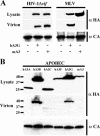
Specific packaging of APOBEC3 proteins into HIV-1 and MLV virions. (A) Analysis of hA3G and mA3 packaging into either Vif-deficient HIV-1 or wild-type MLV virions. A total of 1.5 μg of either pNL4-3-Luc-ΔEnvΔVif or pNCS was transfected into 293T cells together with a plasmid expressing either hA3G-HA or mA3-HA, or the parental plasmid pcDNA3. Producer cell lysates and virion lysates were then subjected to Western analysis using an antibody specific for the HA epitope tag (αHA). Virion lysates were also probed with an antibody specific for the CA protein of either HIV-1 or MLV (αCA), as a loading control. (B) Specific packaging of hA3B but not hA3F into MLV virions. This MLV virion packaging experiment was performed as described for panel A using a range of APOBEC3 expression plasmids.
FIG. 5.
FIG. 5.
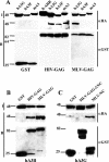
The MLV Gag protein binds to hA3G and hA3B, but not to mA3 or hA3F. (A) 293T cells were cotransfected with plasmids expressing wild-type GST, or GST fused to the full-length HIV-1 or MLV Gag protein, together with plasmids expressing HA-epitope-tagged forms of hA3G, hA3F, or mA3 or the cytoplasmic β-ARR2 protein as a negative control. After cell lysis, GST proteins were collected by incubation with glutathione Sepharose beads and coprecipitated proteins were detected by Western analysis using an antibody specific for the HA-epitope tag (αHA). Controls include Western analysis of the bound beads using a GST-specific antiserum (αGST) and Western analysis of the input cell lysate, prior to incubation with the glutathione beads, using an HA-tag-specific antiserum. (B) Similar to panel A, except that binding of hA3B-HA to GST or to GST fused to full-length HIV-1 or MLV Gag was analyzed. (C) Similar to panel A, except that the binding of hA3G-HA to fusion proteins consisting of GST linked to the MLV Gag NC domain (MLV-NC), or to the MLV Gag polyprotein precisely lacking the carboxy-terminal NC domain (MLV-GAG-ΔNC), was analyzed. I, input proteins; B, bound proteins.
FIG. 6.
FIG. 6.
Binding of hA3G by MLV Gag, and of mA3 by HIV-1 Gag, requires RNA. This analysis was performed as described in Fig. 5, except that the lysates were incubated for 15 min at 37°C, either in the presence or in the absence of RNase A, prior to binding to the glutathione Sepharose beads.

References

    1. Alce, T. M., and W. Popik. 2004. APOBEC3G is incorporated into virus-like particles by a direct interaction with HIV-1 Gag nucleocapsid protein. J. Biol. Chem. 279:34083-34086. - PubMed
    1. Bieniasz, P. D. 2004. Intrinsic immunity: a front-line defense against viral attack. Nat. Immunol. 11:1109-1115. - PubMed
    1. Bishop, K. N., R. K. Holmes, A. M. Sheehy, N. O. Davidson, S.-J. Cho, and M. H. Malim. 2004. Cytidine deamination of retroviral DNA by diverse APOBEC proteins. Curr. Biol. 14:1392-1396. - PubMed
    1. Bogerd, H. P., B. P. Doehle, H. L. Wiegand, and B. R. Cullen. 2004. A single amino acid difference in the host APOBEC3G protein controls the primate species specificity of HIV-1 Vif. Proc. Natl. Acad. Sci. USA 101:3770-3774. - PMC - PubMed
    1. Brownawell, A. M., and I. G. Macara. 2002. Exportin-5, a novel karyopherin, mediates nuclear export of double-stranded RNA binding proteins. J. Cell Biol. 156:53-64. - PMC - PubMed

Publication types

LinkOut - more resources