. 2005 Jul;79(13):8208-16.
doi: 10.1128/JVI.79.13.8208-8216.2005.

Recombinant human immunodeficiency virus type 1 integrase exhibits a capacity for full-site integration in vitro that is comparable to that of purified preintegration complexes from virus-infected cells

Affiliations

Recombinant human immunodeficiency virus type 1 integrase exhibits a capacity for full-site integration in vitro that is comparable to that of purified preintegration complexes from virus-infected cells

Sapna Sinha et al. J Virol. 2005 Jul.

Abstract

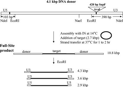
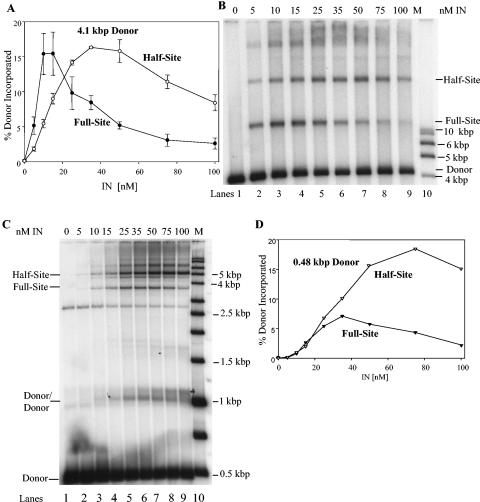
Retrovirus preintegration complexes (PIC) in virus-infected cells contain the linear viral DNA genome (approximately 10 kbp), viral proteins including integrase (IN), and cellular proteins. After transport of the PIC into the nucleus, IN catalyzes the concerted insertion of the two viral DNA ends into the host chromosome. This successful insertion process is termed "full-site integration." Reconstitution of nucleoprotein complexes using recombinant human immunodeficiency virus type 1 (HIV-1) IN and model viral DNA donor substrates (approximately 0.30 to 0.48 kbp in length) that are capable of catalyzing efficient full-site integration has proven difficult. Many of the products are half-site integration reactions where either IN inserts only one end of the viral donor substrate into a circular DNA target or into other donors. In this report, we have purified recombinant HIV-1 IN at pH 6.8 in the presence of MgSO4 that performed full-site integration nearly as efficiently as HIV-1 PIC. The size of the viral DNA substrate was significantly increased to 4.1 kbp, thus allowing for the number of viral DNA ends and the concentrations of IN in the reaction mixtures to be decreased by a factor of approximately 10. In a typical reaction at 37 degrees C, recombinant HIV-1 IN at 5 to 10 nM incorporated 30 to 40% of the input DNA donor into full-site integration products. The synthesis of full-site products continued up to approximately 2 h, comparable to incubation times used with HIV-1 PIC. Approximately 5% of the input donor was incorporated into the circular target producing half-site products with no significant quantities of other integration products produced. DNA sequence analysis of the viral DNA-target junctions derived from wild-type U3 and U5 coupled reactions showed an approximately 70% fidelity for the HIV-1 5-bp host site duplications. Recombinant HIV-1 IN successfully utilized a mutant U5 end containing additional nucleotide extensions for full-site integration demonstrating that IN worked properly under nonideal active substrate conditions. The fidelity of the 5-bp host site duplications was also high with these coupled mutant U5 and wild-type U3 donor ends. These studies suggest that recombinant HIV-1 IN is at least as capable as native IN in virus particles and approaching that observed with HIV-1 PIC for catalyzing full-site integration.


Figures

FIG. 1.
FIG. 1.
Schematic representation of the 4.1-kbp linear DNA substrate, assembly conditions, and strand transfer analyses. The 4.1-kbp donor is depicted (top) with restriction sites and the 3′-OH recessed wt U5 and U3 termini. The donor contained a 420-bp DNA fragment (SupF) that was blunt-end ligated at the AvaI site. The dark line within the rectangular box identifies the approximate length of the LTR regions. IN was assembled with the donor substrate at 14°C followed by the addition of a circular target and strand transfer analysis at 37°C. Two donor ends are inserted by IN into the target producing a 10.8-kbp full-site product. Restriction digestion of the full-site products by EcoRI produced three restriction fragments depicted at the bottom.
FIG. 2.
FIG. 2.
Strand transfer activities of recombinant HIV-1 IN under different assay conditions. (A) IN was assembled at 0, 5, 10, 15, 25, 35, 50, 75, and 100 nM with either 32P-labeled 4.1-kbp or 0.48-kbp donors. The 4.1-kbp donor was assembled with IN for 15 min at 14°C prior to strand transfer for 60 min at 37°C. The 0.48-kbp donor (P2) was assembled with IN for 20 min on ice with DMSO prior to strand transfer for 20 min at 37°C (46). The samples were subjected to 1.5% agarose gel electrophoresis. The dried gels were analyzed by a PhosphorImager, and the percentage of donor incorporated into the integration products was determined from gels shown in panels B and C below. The full-site and half-site products using the 4.1-kbp donors (dark or open circles, respectively) were determined as marked on the figure. The presented data for the 4.1-kbp donor represent triplet measurements with error bar analysis. (B) The full-site and half-site products using the 4.1-kbp donor at various IN concentrations (top) as quantified in panel A. The products and the input donor are indicated on the right. The lane marked “M” on the right contains molecular weight markers as indicated on the right. (C) Same as panel B, except the 0.48-kbp donor was used. The donors/donor products are also shown on the left (46). A nonspecific DNA fragment was present in this P2 DNA preparation that is located in all lanes and migrates just above the 2.5-kbp marker. (D) The percentages of donor incorporated into half-site and full-site products were determined from the data in panel C with the 0.48-kbp donor.
FIG. 3.
FIG. 3.
Time-dependent synthesis of full-site and half-site integration products. (A) IN (5 nM) was preincubated with 32P-labeled 4.1-kbp DNA at 14°C for 15 min. Target DNA was added, and aliquots were removed and processed after 15, 30, 60, 90, 120, 180, and 240 min of incubation at 37°C (lanes 2 to 8, respectively). Lane 1 contained no IN. Lane 9, the 1-kbp DNA ladder markers with several identified on the right. The top two unmarked DNA fragments are 10 and 8 kbp (marked with dots), respectively (see Fig. 2, lane 10). (B) The graph depicts the amounts of full-site and half-site products produced in panel A.
FIG. 4.
FIG. 4.
Assembly properties of IN-DNA complexes capable of full-site integration. (A) IN (5 nM) was preincubated with the wt 32P-labeled 4.1-kbp DNA at 14°C or on ice for 15 min at various NaCl concentrations (bottom). Target DNA was added, and the samples were incubated at 37°C for 120 min. The quantities of integration products produced were determined and plotted against the NaCl concentrations. (B) The same assembly conditions at 14°C were used as described in panel A, except the times of assembly (•, 15 min; ○, 30 min; ▾, 45 min; ▿, 60 min) were different as indicated on the graph. The 3.6-kbp DNA donor was used. In simultaneously performed assays, aliquots were taken and assayed for strand transfer activities at 37°C as indicated at the bottom. The quantities of each product were determined and plotted. The top four lines are full-site products, while the bottom four lines are half-site products.
FIG. 5.
FIG. 5.
Restriction enzyme analysis of strand transfer products produced by HIV-1 IN with the wt 4.1-kbp DNA donor. The strand transfer products were produced by IN at 5 nM (top). The integration products were divided into two nearly equal samples that were not (−) or were (+) digested by EcoRI. Lanes 1 and 2 were DNA donor without IN, while lanes 3 and 4 were with IN. Lane 5, the 1-kbp DNA ladder. The U3 and U5 half-site products (bold) and the 4.3-, 3.6-, and 2.9-kbp full-site products (bold) in lane 4 after EcoRI digestion are identified on the top right and bottom right, respectively. The positions of four DNA markers (not bold) are also illustrated on the right. The marker positioned between the 3.0- and 5.0-kbp markers is 4.0 kbp in length.
FIG. 6.
FIG. 6.
Synthesis of full-site DNA products using a 3.6-kbp donor possessing wt U3 and mutant U5 att ends. (A) The modified U5 att end on the 3.6-kbp donor is described in Fig. 7, row B. Standard reaction conditions were used as described in the legend to Fig. 3 using IN at 5 nM. The graph depicts the amount of full-site and half-site products produced versus time of incubation at 37°C. (B) The U5 end of the 3.6-kbp DNA donor was modified as described in Materials and Methods and as shown in Fig. 7. HIV-1 IN (5 nM) was assembled with labeled donor DNA for 15 min at 14°C. Target DNA was added for strand transfer at 37°C, and the products were removed and processed after 120 min. The products were and were not subjected to EcoRI digestion (top). Lanes 1 and 2 were mutant DNA without IN, while lanes 3 and 4 were with IN. The half-site and full-site products (bold) in lane 4 produced by EcoRI digestion are identified on the top right and bottom right, respectively. The positions of the 1-kbp DNA ladder marker (not bold) are also illustrated on the right.
FIG. 7.
FIG. 7.
HIV-1 mutant U5 att end with wt U3 produced host site duplications with a high fidelity. In row A, the circle junction for the wt U3 and mutant U5 ends in the 3.6-kbp plasmid DNA is shown. Two additional nucleotides (TG) were inserted into U5 and are shown in bold with the rest of the wt att site sequences underlined. The NdeI site at the circle junction was maintained. In row B, the plasmid DNA was digested with NdeI with the additional TG nucleotides (bold) shown. In row C, are shown the full-site integration products produced with wt U3 and mutant U5 ends that were isolated from agarose gels, followed by cloning and sequencing of the donor-target junctions. On the right in this row are shown the number of clones with 0, 5, and 7 bp of target site duplications in which the additional TG dinucleotide was removed prior to integration. In row D are shown clones that had the 5-bp target site sequence duplications but where IN did not remove the TG dinucleotides (bold) prior to producing the full-site products. There were three clones sequenced in this category that showed one each of the 18-, 27-, and 50-bp deletions.

References

    1. Aiyar, A., P. Hindmarsh, A. M. Skalka, and J. Leis. 1996. Concerted integration of linear retroviral DNA by the avian sarcoma virus integrase in vitro: dependence on both long terminal repeat termini. J. Virol. 70:3571-3580. - PMC - PubMed
    1. Anthony, N. J. 2004. HIV-1 integrase: a target for new AIDS chemotherapeutics. Curr. Top. Med. Chem. 4:979-990. - PubMed
    1. Beitzel, B., and F. Bushman. 2003. Construction and analysis of cells lacking the HMGA gene family. Nucleic Acids Res. 31:5025-5032. - PMC - PubMed
    1. Brin, E., and J. Leis. 2002. Changes in the mechanism of DNA integration in vitro induced by base substitutions in the HIV-1 U5 and U3 terminal sequences. J. Biol. Chem. 277:10938-10948. - PubMed
    1. Brin, E., and J. Leis. 2002. HIV-1 integrase interaction with U3 and U5 terminal sequences in vitro defined using substrates with random sequences. J. Biol. Chem. 277:18357-18364. - PMC - PubMed

Publication types

LinkOut - more resources