. 2005 Jul;79(13):8217-29.
doi: 10.1128/JVI.79.13.8217-8229.2005.

An interplay between hypervariable region 1 of the hepatitis C virus E2 glycoprotein, the scavenger receptor BI, and high-density lipoprotein promotes both enhancement of infection and protection against neutralizing antibodies

Affiliations

An interplay between hypervariable region 1 of the hepatitis C virus E2 glycoprotein, the scavenger receptor BI, and high-density lipoprotein promotes both enhancement of infection and protection against neutralizing antibodies

Birke Bartosch et al. J Virol. 2005 Jul.

Abstract

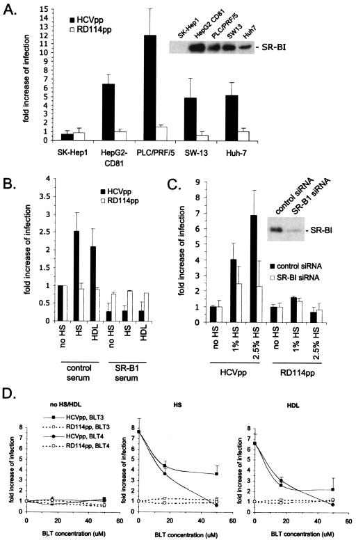
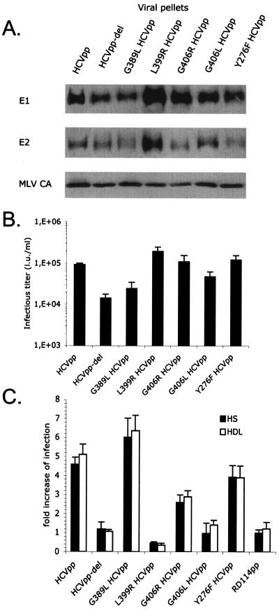
Hepatitis C virus (HCV) circulates in the bloodstream in different forms, including complexes with immunoglobulins and/or lipoproteins. To address the significance of such associations, we produced or treated HCV pseudoparticles (HCVpp), a valid model of HCV cell entry and its inhibition, with naïve or patient-derived sera. We demonstrate that infection of hepatocarcinoma cells by HCVpp is increased more than 10-fold by human serum factors, of which high-density lipoprotein (HDL) is a major component. Infection enhancement requires scavenger receptor BI, a molecule known to mediate HDL uptake into cells as well as HCVpp entry, and involves conserved amino acid positions in hypervariable region 1 (HVR1) of the E2 glycoprotein. Additionally, we show that the interaction with human serum or HDL, but not with low-density lipoprotein, leads to the protection of HCVpp from neutralizing antibodies, including monoclonal antibodies and antibodies present in patient sera. Finally, the deletion or mutation of HVR1 in HCVpp abolishes infection enhancement and leads to increased sensitivity to neutralizing antibodies/sera compared to that of parental HCVpp. Altogether, these results assign to HVR1 new roles which are complementary in helping HCV to survive within its host. Besides immune escape by mutation, HRV1 can mediate the enhancement of cell entry and the protection of virions from neutralizing antibodies. By preserving a balance between these functions, HVR1 may be essential for the viral persistence of HCV.


Figures

FIG. 1.
FIG. 1.
Enhancement of HCVpp infectivity by HS. Infection assays were performed on Huh-7 cells using HCVpp of genotype 1a and RD114pp as the controls. The infectious titers of HCVpp and RD114pp produced in 0.1% FCS were ca. 5 × 10⁴ and 1 × 10⁷ IU/ml, respectively. To obtain viral supernatants containing similar amounts of infectious particles, we therefore diluted all preparations of RD114pp 250-fold. Results show the increases in infection determined by calculating the ratios between the average infectious titers determined in the presence or absence of serum, as indicated. (A) Virions were produced in cell culture media containing the indicated quantities of normal HS or FCS. Results are expressed as ratios between the average infectious titers determined in the presence or absence of serum (mean ± SD, 5). The inset shows a Western blot of purified viral particles that were produced in the absence (−) or presence of 1% HS. The glycoproteins of HCVpp and RD114 were revealed using the A4 and H52 monoclonal antibodies against E1 and E2 (9, 13) and an anti-SU antiserum (ViroMed Biosafety Laboratories), respectively. The MLV capsid (MLV CA) proteins of either pseudoparticle were detected with an anticapsid antiserum (ViroMed Biosafety Laboratories). (B) Virions produced in low-serum medium (0.1% FCS) to which sera from the indicated species were added to 1% were used for infections. The results are expressed as ratios between the average infectious titers determined in the presence of the indicated sera and the titers determined in 0.1% FCS (mean ± SD, 4). (C) Virions were produced in cell culture media containing 0.1% FCS and defined quantities of purified vLDL, LDL, and HDL, as indicated in μg/ml of cholesterol lipoprotein. The results are expressed as ratios between the average infectious titers obtained in the presence or absence of the indicated lipoproteins (mean ± SD, 4). The inset shows a Western blot of purified viral particles produced in low-serum medium supplemented with 6 μg/ml LDL and HDL or not supplemented (−), as indicated. (D) Virions produced in low-serum medium (0.1% FCS) were incubated with defined quantities of vLDL, LDL, and HDL, as indicated in μg/ml, or with defined quantities of an HDL-deficient serum from a patient with Tangier disease (HDL-def. HS) and are compared to normal HS. The results are expressed as ratios between the average infectious titers in the presence of lipoproteins or sera and the titers determined in the absence of lipoproteins or sera.
FIG. 2.
FIG. 2.
Role of SR-BI in facilitation of infection. (A) Results of infection assays with Huh-7, PLC/PRF/5, HepG2-CD81, SW-13, and SK-Hep1 cells that express or do not express SR-BI receptors. HCVpp and RD114pp (1/250 dilution) were produced in cell culture medium containing 0.1% FCS. The infectious titers of HCVpp for these cells in the absence of human serum were as follows: for SK-Hep1, 1.2 × 10³ IU/ml; HepG2-CD81, 1.3 × 10⁴ IU/ml; PLC/PRF/5, 5.7 × 10⁴ IU/ml; SW-13, 103 IU/ml; and Huh-7, 2.4 × 10⁴ IU/ml. Infection assays were performed in the absence or presence of 1% normal HS, which was added during infection. The results are expressed as ratios between the average infectious titers determined in the presence or absence of serum (mean ± SD, 3). The inset shows the expression levels of SR-BI from immunoblotting equal amounts of cell lysates with an SR-BI rabbit antiserum (ab396; Abcam; 1/1,500), as described previously (25). Note that we could detect very small amountsof SR-BI in SK-Hep1 cells on overexposed autoradiographs. (B) HCVpp or RD114pp (1/250 dilution) were produced in cell culture medium containing 0.1% FCS and used in infection assays with Huh-7 cells in the absence of HS (no HS) or in the presence of 1% HS or 6 μg/ml HDL. The same set of infections was performed in the presence of a 1/50-diluted polyclonal anti-SR-BI mouse serum (4). Results are shown as ratios between the average infectious titers determined in the presence or absence of human serum or HDL (mean ± SD, 2). (C) Huh-7 cells expressing a control or anti-SR-BI siRNA (25) were used as target cells for HCVpp or RD114pp (1/250 dilution) produced in 0.1% FCS in the absence (no HS) or presence of HS, as indicated. Downregulation of SR-BI reduced the HCVpp titer fivefold from that of control siRNA-treated Huh-7 cells, as reported previously (25). The inset shows the expression levels of SR-BI from immunoblotting of all lysates, as described previously (25). Results are shown as ratios between the average infectious titers determined in the presence or absence of serum. (D) Huh-7 target cells were treated with 16.7 μM or 50 μM of BLT compounds before and during infection with HCVpp or RD114pp (1/250 dilution) that were produced in 0.1% FCS. As indicated, no HS/HDL, normal HS (2.5%), or HDL (6 μg/ml) was added to the infection reaction mixture. Results are shown as ratios between the average infectious titers determined in the presence or absence of HS or HDL (mean ± SD, 3).
FIG. 3.
FIG. 3.
Role of HVR1 in facilitation of infection. (A) Immunoblots of purified HCVpp generated with E1 and E2 wild-type or mutant glycoproteins (genotype 1a) are shown. E2 point mutations G389L, L399R, G406R, and G406L are located in the HVR1 region. Y276F HCVpp, an E1 point mutant, served as the control. The HCV glycoproteins and the MLV capsid (MLV CA) proteins were revealed with A4 and H52 monoclonal antibodies against E1 and E2 (9, 13) or with an anti-MLV capsid antiserum (ViroMed Biosafety Laboratories). (B) Titers of HCVpp harboring point mutations. Results are expressed as average infectious titers determined on Huh-7 cells in the absence of serum or lipoproteins (mean ± SD, 3). (C) Results of infection of Huh-7 cells with HCVpp or RD114pp produced in 0.1% FCS, with the addition of 2.5% normal HS or 6 μg/ml HDL to the infection reaction mixture. The concentrations of viral supernatants were adjusted to obtain infection of ca. 5 to 10% of target cells. The results are expressed as ratios between average infectious titers determined in the presence or absence of serum or lipoproteins (mean ± SD, 3).
FIG. 4.
FIG. 4.
Human serum or HDL protects HCVpp from neutralization. (A) Effects of sera from a cohort of acutely infected HCV patients (24) on infectivity of the indicated HCVpp. Data were obtained over a time interval of 16 weeks and are shown for four patients who are representative of the cohort. The HCV RNA kinetics for each patient were measured weekly after inclusion in the cohort (HCV RNAs [given in IU/ml], analyzed by means of a third-generation, branched-DNA-based assay [Versant HCV RNA 3.0 assay; Bayer Diagnostics, Tarrytown, N.J.]). The 13-patient cohort consisted of two groups. The first group (7/13 patients), represented by patients 3 and 4 (Pt-3 and Pt-4), showed a >3- to 4-log reduction in HCV RNA titers in the second half of the study period (24). Patients 8 and 9 (Pt-8 and Pt-9) represent the second group (6/13 patients), in whom replication levels remained high (<1-log decrease in HCV RNA titers) throughout the entire study period (24). Serum samples chosen from the beginning, middle, and end of the study period from these four patients were investigated in neutralization assays at 1/50 dilutions with HCVpp (104 IU) of genotype 1b, strain CG1b (HCVpp-1b); HCVpp of genotype 1a, strain H77 (HCVpp-1a); or their HVR1-deleted counterparts (HCVpp-del1b and HCVpp-del1a, respectively) by incubation for 30 min at room temperature before infection of Huh-7 target cells. The results are expressed as mean percentages (±standard deviations [SD]; n = 3) of inhibition of the infectious titers relative to inhibition from incubation with medium devoid of patient sera. Note that no or poor neutralization was detected in Pt-3 and Pt-4 (group 1) with HCVpp-1a, and no neutralization at all was detected in Pt-8 and Pt-9 (group 2) with both HCVpp-1a and HCVpp-1b (Table 2). The specificity of neutralization was controlled with RD114pp, against which no antibodies were detected in HS (3). Nonspecific inhibition of RD114pp (data not shown) over a value of ±20% was never detected. (B) Titration of neutralizing antibodies in total IgG purified from chronically infected HCV patients. Neutralization assays were performed in the absence of HS (no HS) or in the presence of 2.5% HS, 39 μg/ml LDL, or 6 μg/ml HDL. The results are expressed as mean percentages (±SD; n = 4) of inhibition of the infectious titers relative to inhibition from incubation with medium devoid of antibodies. (C) Neutralization curves of the AP33 monoclonal HCV E2 antibody in the absence (no HS) or presence of 2.5% HS using HCVpp harboring the indicated point mutations in HVR1. The results are expressed as the mean percentages (±SD; n = 3) of inhibition of the infectious titers relative to incubation with medium devoid of antibodies. ID50 values are indicated by dotted lines.

References

    1. Agnello, V., G. Ábel, M. Elfahal, G. B. Knight, and Q.-X. Zhang. 1999. Hepatitis C virus and other Flaviviridae viruses enter cells via low density lipoprotein receptor. Proc. Natl. Acad. Sci. USA 96:12766-12771. - PMC - PubMed
    1. Bartosch, B., J. Bukh, J. C. Meunier, C. Granier, R. E. Engle, W. C. Blackwelder, S. U. Emerson, F. L. Cosset, and R. H. Purcell. 2003. In vitro assay for neutralizing antibody to hepatitis C virus: evidence for broadly conserved neutralization epitopes. Proc. Natl. Acad. Sci. USA 100:14199-14204. - PMC - PubMed
    1. Bartosch, B., J. Dubuisson, and F. L. Cosset. 2003. Infectious hepatitis C virus pseudo-particles containing functional E1-E2 envelope protein complexes. J. Exp. Med. 197:633-642. - PMC - PubMed
    1. Bartosch, B., A. Vitelli, C. Granier, C. Goujon, J. Dubuisson, S. Pascale, E. Scarselli, R. Cortese, A. Nicosia, and F.-L. Cosset. 2003. Cell entry of hepatitis C virus requires a set of co-receptors that include the CD81 tetraspanin and the SR-B1 scavenger receptor. J. Biol. Chem. 278:41624-41630. - PubMed
    1. Booth, J. C., U. Kumar, D. Webster, J. Monjardino, and H. C. Thomas. 1998. Comparison of the rate of sequence variation in the hypervariable region of E2/NS1 region of hepatitis C virus in normal and hypogammaglobulinemic patients. Hepatology 27:223-227. - PubMed

Publication types

Substances

LinkOut - more resources