. 2005 Jul;79(13):8230-6.
doi: 10.1128/JVI.79.13.8230-8236.2005.

New DNA viruses identified in patients with acute viral infection syndrome

Affiliations

New DNA viruses identified in patients with acute viral infection syndrome

Morris S Jones et al. J Virol. 2005 Jul.

Abstract

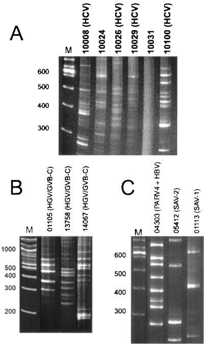
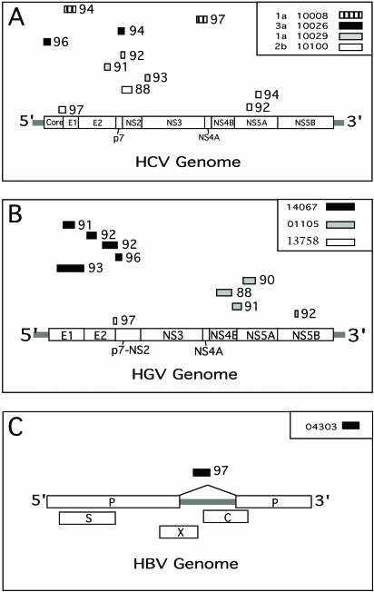
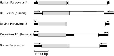
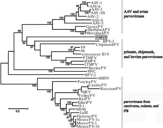
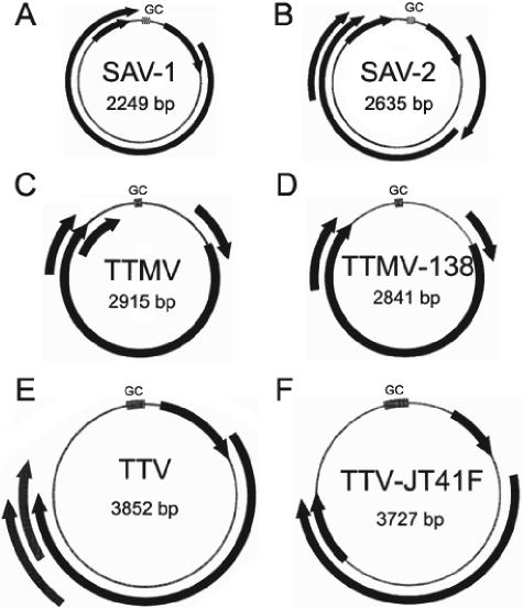
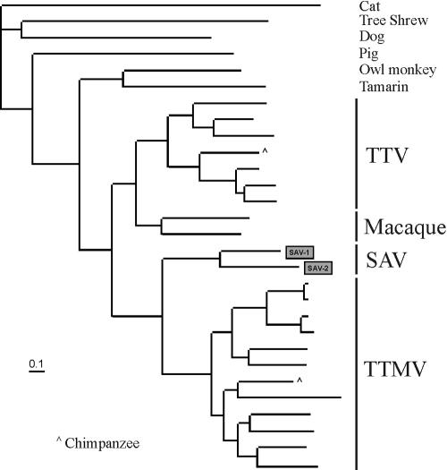
A sequence-independent PCR amplification method was used to identify viral nucleic acids in the plasma samples of 25 individuals presenting with symptoms of acute viral infection following high-risk behavior for human immunodeficiency virus type 1 transmission. GB virus C/hepatitis G virus was identified in three individuals and hepatitis B virus in one individual. Three previously undescribed DNA viruses were also detected, a parvovirus and two viruses related to TT virus (TTV). Nucleic acids in human plasma that were distantly related to bacterial sequences or with no detectable similarities to known sequences were also found. Nearly complete viral genome sequencing and phylogenetic analysis confirmed the presence of a new parvovirus distinct from known human and animal parvoviruses and of two related TTV-like viruses highly divergent from both the TTV and TTV-like minivirus groups. The detection of two previously undescribed viral species in a small group of individuals presenting acute viral syndrome with unknown etiology indicates that a rich yield of new human viruses may be readily identifiable using simple methods of sequence-independent nucleic acid amplification and limited sequencing.


Figures

FIG. 1.
FIG. 1.
DNase-SISPA amplification products from (A) six HCV-positive plasma samples used to test methodology, (B) three RNA-extracted plasma samples, and (C) three DNA-extracted plasma samples. PCR products were analyzed on a 6.5% polyacrylamide gel. Patient identification numbers are shown over the lanes. Viral sequences identified are shown in parentheses. Lanes M contain molecular weight markers in base pairs.
FIG. 2.
FIG. 2.
Genomic locations of subcloned viral sequences homologous to HCV (A), GBV-C/HGV (B), and HBV (C). Patient identification numbers (and HCV genotypes in panel A) are indicated in the symbol key, and viral subtypes are indicated for HCV sequences. Nucleotide percent similarity values are indicated adjacent to the subcloned fragments.
FIG. 3.
FIG. 3.
Genetic organization of PARV4 compared to those of B19, BPV-3, parvovirus H1, and goose parvoviruses. The gray and white boxes represent the genes encoding for nonstructural and structural proteins, respectively. The arrows indicate the positions of the terminal repeat sequences. The arrows at the extremities of parvovirus H1 denote that the terminal repeat sequences are dissimilar.
FIG. 4.
FIG. 4.
Phylogenetic analysis of the PARV4 genome and other members of the Parvoviridae subfamily. Abbreviations: Barbduck PV, Barbary duck parvovirus; MuscduckPV, Muscovy duck parvovirus; PTMPV, pig-tailed macaque parvovirus; RMPV, rhesus macaque parvovirus; LTMPV, long-tailed macaque parvovirus; MVC, minute virus of canines; AMDV, Aleutian mink disease virus; MEV, mink enteritis virus; Kilrat, Kilham rat parvovirus; MVM, minute virus of mice.
FIG. 5.
FIG. 5.
Genetic organization of (A) SAV-1, (B) SAV-2, (C) TTMV, (D) TTMV-238, (E) TTV, and (F) TTV-JT41F. Arrows represent ORFs detected in each virus. The GC-rich region (GC) has a GC content greater than 72%. ORFinder (National Center for Biotechnology Information) was used to determine the ORFs for each virus as described in Materials and Methods.
FIG. 6.
FIG. 6.
Phylogenetic analysis of the large ORFs of SAV, TTV, and TTMV found in a range of mammalian species. All viral sequences originate from humans unless indicated otherwise.

References

    1. Allander, T., S. U. Emerson, R. E. Engle, R. H. Purcell, and J. Bukh. 2001. A virus discovery method incorporating DNase treatment and its application to the identification of two bovine parvovirus species. Proc. Natl. Acad. Sci. USA 98:11609-11614. - PMC - PubMed
    1. Baylis, S. A., N. Shah, and P. D. Minor. 2004. Evaluation of different assays for the detection of parvovirus B19 DNA in human plasma. J. Virol. Methods 121:7-16. - PubMed
    1. Breitbart, M., I. Hewson, B. Felts, J. M. Mahaffy, J. Nulton, P. Salamon, and F. Rohwer. 2003. Metagenomic analyses of an uncultured viral community from human feces. J. Bacteriol. 185:6220-6223. - PMC - PubMed
    1. Breitbart, M., P. Salamon, B. Andresen, J. M. Mahaffy, A. M. Segall, D. Mead, F. Azam, and F. Rohwer. 2002. Genomic analysis of uncultured marine viral communities. Proc. Natl. Acad. Sci. USA 99:14250-14255. - PMC - PubMed
    1. Burnouf, T., and M. Radosevich. 2000. Reducing the risk of infection from plasma products: specific preventative strategies. Blood Rev. 14:94-110. - PubMed

Publication types

MeSH terms

LinkOut - more resources