Comparative Study
. 2005 Aug;170(4):1747-59.
doi: 10.1534/genetics.105.041749. Epub 2005 Jun 14.

Adult myogenesis in Drosophila melanogaster can proceed independently of myocyte enhancer factor-2

Affiliations
Comparative Study

Adult myogenesis in Drosophila melanogaster can proceed independently of myocyte enhancer factor-2

Phillip W Baker et al. Genetics. 2005 Aug.

Abstract

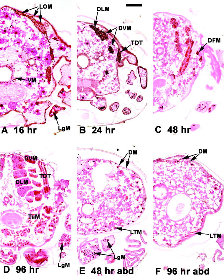
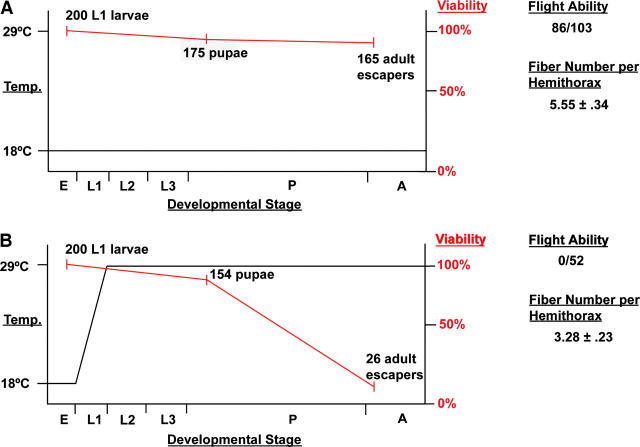
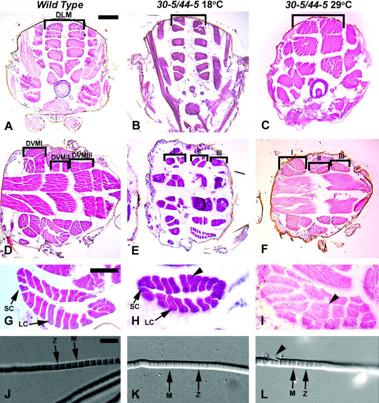
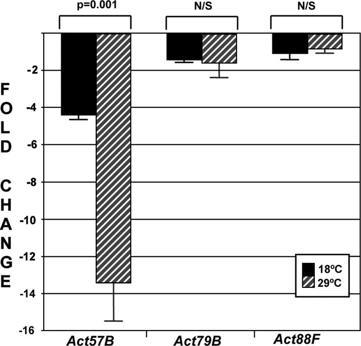
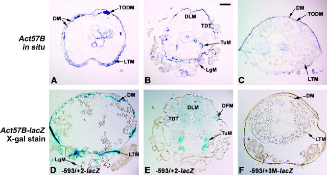
Myocyte enhancer factor-2 (MEF2) is a transcription factor that is necessary for embryonic muscle development in Drosophila and vertebrates; however, whether this factor is required during later muscle development remains largely unknown. Using heteroallelic combinations of different Mef2 mutant alleles, we isolated and characterized a temperature-sensitive combination. Through temperature-shift experiments, we obtained adult animals that were lacking proper MEF2 function. Many of these individuals died as mature pupae, and those that eclosed showed poor locomotion and an inability to fly. Histological analysis of these animals revealed a requirement for MEF2 in skeletal muscle patterning, although these animals had strikingly normal amounts of muscle tissue. Using quantitative polymerase chain reaction, we determined that expression of the MEF2-regulated actin gene Act57B was severely reduced in these animals. By contrast myofibrillar actin genes unique to the adult stage were only mildly affected. Since MEF2 mutant adults were still capable of forming muscle tissue, we conclude that MEF2 is required for the expression of only a subset of muscle structural genes in the adult. These results indicate that additional muscle-specific factors function to control the myogenesis of complex and diverse muscle in the adult.


Figures

F<sc>igure</sc> 1.—
Figure 1.—
Expression of Mef2 during adult skeletal muscle development. Transverse paraffin sections (with dorsal side up) of aged pupae were reacted with a MEF2 antibody (brown stain) and counterstained with eosin. (A–D) Thoracic sections. (E and F) Abdominal sections (abd). Half-sections are shown for detail. (A) At 16 hr APF, myoblasts from the wing disc migrated dorsally to surround the LOM, which form the templates for the DLMs. Myoblasts also migrated ventrally. Myoblasts in the forming leg (LgM) were also observed. (B) At 24 hr APF MEF2-positive myoblasts had coalesced to form the major IFM fibers, the DLMs, and the DVMs. The developing tergal depressor of the trochanter (TDT) was also observed at this stage. (C) At 48 hr APF Mef2 expression persisted in the major fibers and was observed in direct flight muscles (DFMs) apposed to the base of the wing. (D) Mef2 expression persisted to the end of pupal development in all thoracic muscle types, including the fibrillar DLM and DVM and the TDT and other tubular muscles (TuM). (E) Mef2 expression was also detected in the developing abdominal muscles throughout pupal development, although this was more apparent at midpupal stages in the dorsal muscles (DM) and the lateral transverse muscles (LTM). (F) MEF2 was also detected in mature abdominal muscles. MEF2 was also detected in various muscles surrounding the gut (VM). Bar, 100 μm.
F<sc>igure</sc> 2.—
Figure 2.—
Muscle development in wild-type and Mef2 mutant embryos. Embryos at stage 16 were stained in parallel with an anti-myosin heavy-chain (MHC) antibody. (A) Wild-type animals showed a large number of MHC-positive body wall muscle fibers. (A′) The lateral transverse (LT) muscles 1–3 are indicated (arrowheads). (B) 30-5/44-5 mutants raised at 18° showed numerous muscle fibers although slightly reduced in number and with a reduced staining intensity. (Inset) The dorsal vessel formed in these mutants, but commonly showed organizational defects (arrowhead). (B′) The number of LT fibers was reduced. (C) 30-5/44-5 mutants raised at 29° showed a massively reduced number of muscle fibers. (C′) LT 1-3 were absent or reduced in number in all segments. (D) Mef222-21 mutant homozygotes showed a failure of muscle formation. The phenotypes of both wild-type and mutant genotypes were similar within each genotype stained. Anterior is to the left and dorsal is top. Inset in B is a dorsal view. Bar, 100 μm for A–D; 50 μm for A′–D′.
F<sc>igure</sc> 3.—
Figure 3.—
Summary of temperature-shift experiments and results obtained. The left axes denote temperature treatment of 30-5/44-5 mutants (black line). Viability of these mutants starting with 100% at L1 is represented by the red line and listed on the right axes. Indicated to the right of the graphs are the flight ability and the number of DLM fibers per hemithorax in adult escapers for each treatment. (A) Mutants raised at 18° throughout development. (B) Mutants raised at 18° during embryogenesis and at 29° thereafter. Developmental stages: (E) embryo, (L1–3) larval instars, (P) pupa, (A) adult.
F<sc>igure</sc> 4.—
Figure 4.—
Histological analyses of wild-type and 30-5/44-5 mutants raised at the permissive and restrictive temperatures. Wild-type animals are 96-hr APF, and mutant animals raised at 18° and 29° were of equivalent stages. (A–C) Transverse thoracic sections to visualize DLMs (bracketed), dorsal to top. (D–F) Horizontal thoracic sections to visualize the DVM I-III fibers (bracketed), anterior to the left. (G–I) Horizontal view of TDT muscle, anterior to left. (J–L), Dissected myofibrils. Sections in A–I were stained with hematoxylin and eosin. (A) Wild type. (B) 30-5/44-5 mutant raised at 18°, where DLM fiber number and arrangement were similar to wild type. (C) 30-5/44-5 mutant raised at 29°, note the reduced number of DLM fibers. (D) Wild type. (E) 30-5/44-5 mutant raised at 18°. DVM fiber number remained similar to wild type but some patterning defects were seen. (F) 30-5/44-5 mutant raised at 29°, fiber number was not notably different from permissive temperature. (G) Wild-type TDT, indicating the small cells (SC) and the large cells (LC) of the muscle. (H) TDT of a 30-5/44-5 mutant raised at 18°, showing a nearly normal structure, but with a single abnormally shaped large cell (arrowhead). (I) TDT of a 30-5/44-5 mutant raised at 29°: the central lumen characteristic of this muscle was missing and many of the fibers were shaped oddly. (J) Myofibril isolated from the DLM of wild-type animal. Note the regular structure of the sarcomere and prominent Z-lines and M-lines (arrows). (K) Myofibril isolated from a DLMof 30-5/44-5 animal raised at 18° was apparently normal. (L) Myofibril isolated from a DLM of 30-5/44-5 animal raised at 29°; note the frayed appearance of the fiber at left (arrowhead). Bar for A–F, 100 μm; for G–I, 50 μm; for J–L, 5 μm.
F<sc>igure</sc> 5.—
Figure 5.—
QPCR analysis of muscle gene expression in mutant adults. The transcript levels of three actin genes, Act57B, Act79B, and Act88F, were determined from total RNA of mature pupae. Fold expression changes were calculated for 30-5/44-5 at 18° (solid bars) and at 29° (shaded bars), compared to wild type. Values represent the average of four assays, and error bars represent the standard error of the mean. Act57B expression was dramatically reduced in the 30-5/44-5 mutant animals as compared to the other actin genes assayed. Student's t-test indicated that the differences in Act79B and Act88F transcript levels in permissive or restrictive mutants were not significant (N/S), but that the change in Act57B expression was highly significant.
F<sc>igure</sc> 6.—
Figure 6.—
Expression and regulation of Act57B in adult Drosophila. (A–C) In situ hybridization to transverse paraffin sections (with dorsal side up) using either a 3′-UTR antisense Act57B probe (A and B) or a sense control probe (C). (A) In the abdomen, Act57B expression was detected in the DM, including the temporary oblique dorsal muscles (TODM) which are used for eclosion, as well as in the ventrally located LTM. (B) In the thorax, Act57B was detected in a small subset of TuM and in the leg (LgM) but not in the TDT or the IFMs. (C) No reproducible signal was observed with the control probe. (D–F) Transverse cryosections of Act57B-lacZ transgenic animals stained with X-gal. (D) The 593/+2 Act57B-lacZ transgene was active in the abdomen in the same muscle cells as the endogenous gene. (E) Transgene expression was also observed in the thoracic tubular muscles, although additionally at low levels in the IFMs. This may represent leakiness of the transgene or possibly a weak early expression of Act57B in these muscles. (F) The 593/+2 transgene in which the MEF2 site was mutated was inactive in adult sections. Bar, 100 μm.

References

    1. Anant, S., S. Roy and K. Vijayraghavan, 1998. Twist and Notch negatively regulate adult muscle differentiation in Drosophila. Development 125: 1361–1369. - PubMed
    1. Andres, V., M. Cervera and V. Mahdavi, 1995. Determination of the consensus binding site for MEF2 expressed in muscle and brain reveals tissue-specific sequence constraints. J. Biol. Chem. 270: 23246–23249. - PubMed
    1. Arredondo, J. J., R. M. Ferreres, M. Maroto, R. M. Cripps, R. Marco et al., 2001. Control of Drosophila Paramyosin/Miniparamyosin gene expression: differential regulatory mechanisms for muscle-specific transcription. J. Biol. Chem. 276: 8278–8287. - PubMed
    1. Ashburner, M., 1989 Drosophila: A Laboratory Handbook. Cold Spring Harbor Laboratory Press, Cold Spring Harbor, NY.
    1. Bagni, C., S. Bray, J. A. Gogos, F. C. Kafatos and T. Hsu, 2002. The Drosophila zinc finger transcription factor CF2 is a myogenic marker downstream of MEF2 during muscle development. Mech. Dev. 117: 265–268. - PubMed

Publication types

MeSH terms

Substances

LinkOut - more resources