TCR Valpha14 natural killer T cells function as effector T cells in mice with collagen-induced arthritis
- PMID: 15958069
- PMCID: PMC1809413
- DOI: 10.1111/j.1365-2249.2005.02817.x
TCR Valpha14 natural killer T cells function as effector T cells in mice with collagen-induced arthritis
Abstract
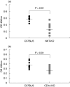
Natural killer (NK) T cells are a unique, recently identified cell population and are suggested to act as regulatory cells in autoimmune disorders. In the present study, designed to investigate the role of NKT cells in arthritis development, we attempted to induce arthritis by immunization of type II collagen (CIA) in Jalpha281 knock out (NKT-KO) and CD1d knock out (CD1d-KO) mice, which are depleted of NKT cells. From the results, the incidence of arthritis (40%) and the arthritis score (1.5 +/- 2.2 and 2.0 +/- 2.7) were reduced in NKT-KO and CD1d-KO mice compared to those in respective wild type mice (90%, 5.4 +/- 3.2 and 2.0 +/- 2.7, P < 0.01). Anti-CII antibody levels in the sera of NKT-KO and CD1d-KO mice were significantly decreased compared to the controls (OD values; 0.32 +/- 0.16 and 0.29 +/- 0.06 versus 0.58 +/- 0.08 and 0.38 +/- 0.08, P < 0.01). These results suggest that NKT cells play a role as effector T cells in CIA. Although the cell proliferative response and cytokine production in NKT-KO mice after the primary immunization were comparable to those in wild type mice, the ratios of both activated T or B cells were lower in NKT-KO mice than wild type mice after secondary immunization (T cells: 9.9 +/- 1.8% versus 16.0 +/- 3.4%, P < 0.01, B cells: 4.1 +/- 0.5% versus 5.1 +/- 0.7%, P < 0.05), suggesting that inv-NKT cells contribute to the pathogenicity in the development phase of arthritis. In addition, IL-4 and IL-1beta mRNA expression levels in the spleen during the arthritis development phase were lower in NKT-KO mice, while the IFN-gamma mRNA expression level was temporarily higher. These results suggest that inv-NKT cells influence cytokine production in arthritis development. In conclusion, inv-NKT cells may promote the generation of arthritis, especially during the development rather than the initiation phase.
Figures


Similar articles
-
The involvement of V(alpha)14 natural killer T cells in the pathogenesis of arthritis in murine models.Arthritis Rheum. 2005 Jun;52(6):1941-8. doi: 10.1002/art.21056. Arthritis Rheum. 2005. PMID: 15934073
-
A novel function of Valpha14+CD4+NKT cells: stimulation of IL-12 production by antigen-presenting cells in the innate immune system.J Immunol. 1999 Jul 1;163(1):93-101. J Immunol. 1999. PMID: 10384104
-
Collagen type II (CII)-specific antibodies induce arthritis in the absence of T or B cells but the arthritis progression is enhanced by CII-reactive T cells.Arthritis Res Ther. 2004;6(6):R544-50. doi: 10.1186/ar1217. Epub 2004 Sep 23. Arthritis Res Ther. 2004. PMID: 15535832 Free PMC article.
-
The regulatory role of Valpha14 NKT cells in innate and acquired immune response.Annu Rev Immunol. 2003;21:483-513. doi: 10.1146/annurev.immunol.21.120601.141057. Epub 2001 Dec 19. Annu Rev Immunol. 2003. PMID: 12543936 Review.
-
Natural killer T cells and rheumatoid arthritis: friend or foe?Arthritis Res Ther. 2005;7(2):88-9. doi: 10.1186/ar1714. Epub 2005 Feb 14. Arthritis Res Ther. 2005. PMID: 15743495 Free PMC article. Review. No abstract available.
Cited by
-
Diverse cytokine production by NKT cell subsets and identification of an IL-17-producing CD4-NK1.1- NKT cell population.Proc Natl Acad Sci U S A. 2008 Aug 12;105(32):11287-92. doi: 10.1073/pnas.0801631105. Epub 2008 Aug 6. Proc Natl Acad Sci U S A. 2008. PMID: 18685112 Free PMC article.
-
It Takes "Guts" to Cause Joint Inflammation: Role of Innate-Like T Cells.Front Immunol. 2018 Jun 29;9:1489. doi: 10.3389/fimmu.2018.01489. eCollection 2018. Front Immunol. 2018. PMID: 30008717 Free PMC article. Review.
-
Specific Inhibition of Soluble γc Receptor Attenuates Collagen-Induced Arthritis by Modulating the Inflammatory T Cell Responses.Front Immunol. 2019 Feb 8;10:209. doi: 10.3389/fimmu.2019.00209. eCollection 2019. Front Immunol. 2019. PMID: 30800133 Free PMC article.
-
Activation of natural killer T cells by α-carba-GalCer (RCAI-56), a novel synthetic glycolipid ligand, suppresses murine collagen-induced arthritis.Clin Exp Immunol. 2011 May;164(2):236-47. doi: 10.1111/j.1365-2249.2011.04369.x. Epub 2011 Mar 10. Clin Exp Immunol. 2011. PMID: 21391989 Free PMC article.
-
The requirement of natural killer T-cells in tolerogenic APCs-mediated suppression of collagen-induced arthritis.Exp Mol Med. 2010 Aug 31;42(8):547-54. doi: 10.3858/emm.2010.42.8.055. Exp Mol Med. 2010. PMID: 20610917 Free PMC article.
References
-
- Sumida T, Maeda T, Taniguchi M, Nishioka K. TCRAV24 gene expression in double negative T cells in systemic lupus erythematosus. Lupus. 1998;7:565–8. - PubMed
-
- Maeda T, Keino H, Asahara H, et al. Decreased TCR AV24AJ18+ double negative T cells in rheumatoid synovium. Rheumatology. 1999;38:186–8. - PubMed
-
- Sharif S, Arreaza GA, Zucker P, Denovitch TL. Regulatory natural killer T cells protect against spontaneous and recurrent type I diabetes. Ann NY Acad Sci. 2002;958:77–88. - PubMed
MeSH terms
Substances
LinkOut - more resources
Full Text Sources
Other Literature Sources
Molecular Biology Databases
Research Materials

