

Review
doi: 10.1136/hrt.2004.050443.
Clinical applications of intracardiac echocardiography in interventional procedures
Affiliations
- PMID: 15958380
- PMCID: PMC1768980
- DOI: 10.1136/hrt.2004.050443
Item in Clipboard
Review
Clinical applications of intracardiac echocardiography in interventional procedures
Heart.
2005 Jul.
No abstract available
Figures
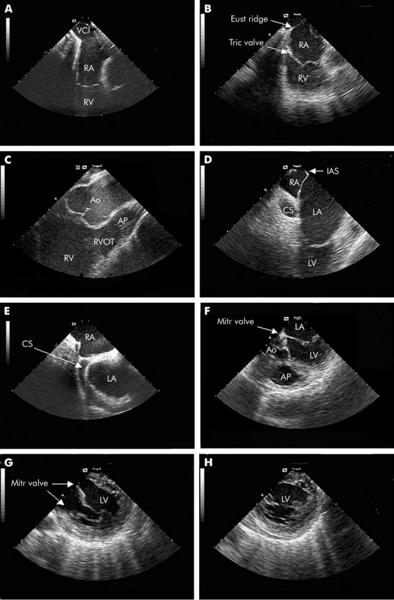
Different views obtained with the intracardiac echocardiography (ICE) transducer in either the right atrium or the right ventricle. See text for explanation of panels A–H. Ao, aorta; AP, pulmonary artery; CS, coronary sinus; Eust ridge, Eustachian ridge; IAS, interatrial septum; LA, left atrium; LV, left ventricle; Mitr valve, mitral valve; RA, right atrium; RV, right ventricle; RVOT, right ventricular outflow tract; Tric valve, tricuspid valve; VCI, inferior caval vein.

(A) Spontaneous contrast in the left atrial appendage (LAA), a risk factor for thrombus formation. (B) Function of the LAA can be determined with phased array ICE, using pulsed wave Doppler. (C) Thrombus in the LAA. (D) The Valsalva manoeuvre is performed before transseptal puncture to determine the presence of patent foramen ovale. Contrast bubbles can be observed in the right atrium (arrow). The lack of bubbles in the left atrium confirms the presence of a closed foramen. (E) The catheter is shifted through the septum through a patent foramen ovale (PFO). (F) Tenting of the septum caused by stable contact of the transseptal dilator/needle. LA, left atrium; RA: right atrium.

Closure of an atrial septal defect (ASD) under ICE guidance. (A) Colour Doppler demonstrating an ASD located in the inferior part of the atrial septum. (B) Relation of the ASD (arrow) to intracardiac structures. (C) Positioning of the Amplatzer closure device. (D) After several minutes, flattening of the closure device occurs. The device is positioned stable in between the right and left atria. Ao, aorta; LA, left atrium; RA, right atrium; RV, right ventricle.

(A) Left PVs. There is a common ostium of the left PVs (diameter 2.04 cm). (B) Colour Doppler in the right PVs. Three right PVs are visible. (C) Ablation catheter at ostium of the left superior PV. Note the presence of microbubbles, indicating heat loss during ablation. (D) Pulsed wave Doppler measurements of flow in the PVs. LA, left atrium; LIPV, left inferior pulmonary vein; LSPV, left superior pulmonary vein; RA, right atrium; RSPV, right superior pulmonary vein; RMPV, right middle pulmonary vein; RIPV, right inferior pulmonary vein.

(A) The cavo-tricuspid isthmus (double arrow) demonstrated with ICE as the area in between the inferior caval vein and the tricuspid annulus. (B) Imaging of the cavo-tricuspid isthmus area in a patient with atrial flutter and Ebstein anomaly. Due to the low insertion of the tricuspid valve leaflet, part of the right ventricle is incorporated in the right atrium (A indicates “atrialised” right ventricle). Ao, aorta; PA, pulmonary artery; Tric valve, tricuspid valve; RA, right atrium; RV, right ventricle.

(A) Long axis view of the left ventricle (LV) and an apical LV aneurysm (aneur) in a patient with ventricular tachycardia. (B) Ablation catheter at the site of the LV aneurysm (short axis view), demonstrating perpendicular catheter tip–tissue contact. (C) Postero-basal LV aneurysm. (D) Microbubble formation during ablation, indicating heat loss. cath tip, catheter tip.

(A) Position of catheters as seen on fluoroscopy in a patient with ventricular tachycardia originating from the right ventricular outflow tract (RVOT). (B) ICE image of the same patient, demonstrating the position of the ablation catheter in the RVOT. (C) Electro-anatomical map of the right ventricle (RV) (CARTO). The activation of the RV is colour encoded. Red indicates the area of earliest activation in the RVOT. (D) Imaging in a patient with ventricular tachycardia secondary to ARVD/C. An aneurysmatic area was observed at the inferior part of the RV. Early fragmented signals were recorded at this site during tachycardia and subsequent radiofrequency catheter ablation was initiated at this site. aneur, aneurysm; Ao, aorta; AP, pulmonary artery; cath tip, catheter tip; ICE, intracardiac ultrasound catheter; RA, right atrium; RVA, right ventricular apex.

(A) Evaluation of an intracardiac mass by ICE. An extensive mass is present in the right atrium (RA), which extends over the tricuspid annulus to the wall of the right ventricle (RV). (B) Biopsies were taken under ICE guidance. The final diagnosis was B cell non-Hodgkin lymphoma.
References
-
- Dairywala IT, Li P, Liu Z, et al. Catheter-based interventions guided solely by a new phased-array intracardiac imaging catheter: in vivo experimental studies. J Am Soc Echocardiogr 2002;15:150–8. - PubMed
-
- Chu E, Fitzpatrick AP, Chin MC, et al. Radiofrequency catheter ablation guided by intracardiac echocardiography. Circulation 1994;89:1301–5. - PubMed
-
- Antonielli E, Pizzuti A, Palinkas A, et al. Clinical value of left atrial appendage flow for prediction of long-term sinus rhythm maintenance in patients with nonvalvular atrial fibrillation. J Am Coll Cardiol 2002;39:1443–9. - PubMed
-
- Morton JB, Sanders P, Sparks PB, et al. Usefulness of phased-array intracardiac echocardiography for the assessment of left atrial mechanical “stunning” in atrial flutter and comparison with multiplane transesophageal echocardiography. Am J Cardiol 2002;90:741–6. ▸ This paper demonstrates that phased array ICE can be a good alternative to TOE for the evaluation of left atrial mechanical function. - PubMed
-
- Thakur RK, Klein GJ, Yee R, et al. Embolic complications after radiofrequency catheter ablation. Am J Cardiol 1994;74:278–9. - PubMed
Publication types
MeSH terms
LinkOut - more resources
Full Text Sources
Other Literature Sources