. 2005 Aug;16(8):3896-907.
doi: 10.1091/mbc.e05-02-0118. Epub 2005 Jun 15.

Cell cycle-dependent dynamics and regulation of mitotic kinesins in Drosophila S2 cells

Affiliations

Cell cycle-dependent dynamics and regulation of mitotic kinesins in Drosophila S2 cells

Gohta Goshima et al. Mol Biol Cell. 2005 Aug.

Abstract

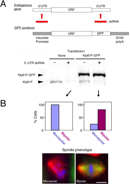
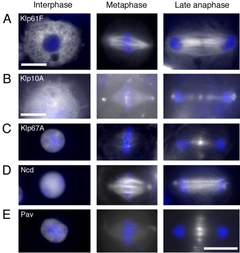
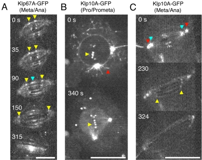
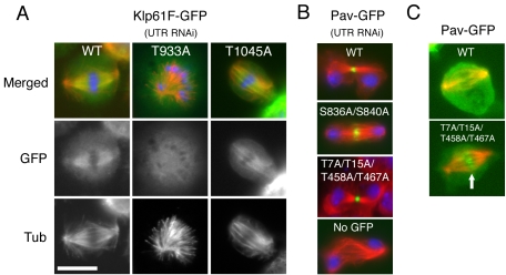
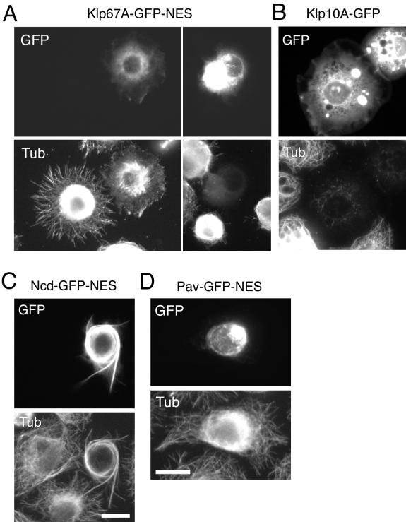
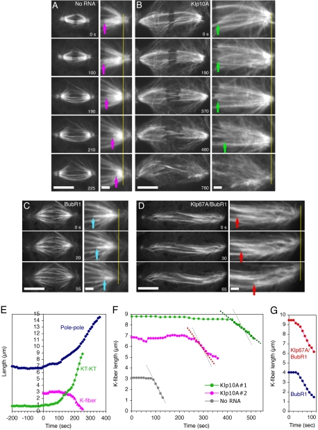
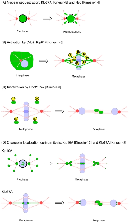
Constructing a mitotic spindle requires the coordinated actions of several kinesin motor proteins. Here, we have visualized the dynamics of five green fluorescent protein (GFP)-tagged mitotic kinesins (class 5, 6, 8, 13, and 14) in live Drosophila Schneider cell line (S2), after first demonstrating that the GFP-tag does not interfere with the mitotic functions of these kinesins using an RNA interference (RNAi)-based rescue strategy. Class 8 (Klp67A) and class 14 (Ncd) kinesin are sequestered in an active form in the nucleus during interphase and engage their microtubule targets upon nuclear envelope breakdown (NEB). Relocalization of Klp67A to the cytoplasm using a nuclear export signal resulted in the disassembly of the interphase microtubule array, providing support for the hypothesis that this kinesin class possesses microtubule-destabilizing activity. The interactions of Kinesin-5 (Klp61F) and -6 (Pavarotti) with microtubules, on the other hand, are activated and inactivated by Cdc2 phosphorylation, respectively, as shown by examining localization after mutating Cdc2 consensus sites. The actions of microtubule-destabilizing kinesins (class 8 and 13 [Klp10A]) seem to be controlled by cell cycle-dependent changes in their localizations. Klp10A, concentrated on microtubule plus ends in interphase and prophase, relocalizes to centromeres and spindle poles upon NEB and remains at these sites throughout anaphase. Consistent with this localization, RNAi analysis showed that this kinesin contributes to chromosome-to-pole movement during anaphase A. Klp67A also becomes kinetochore associated upon NEB, but the majority of the population relocalizes to the central spindle by the timing of anaphase A onset, consistent with our RNAi result showing no effect of depleting this motor on anaphase A. These results reveal a diverse spectrum of regulatory mechanisms for controlling the localization and function of five mitotic kinesins at different stages of the cell cycle.


Figures

Figure 1.
Figure 1.
Rescue assay in S2 cells. (A) Top, schematic representation of a rescue assay in S2 cells. dsRNA targeting 5′UTR or 3′UTR is used for RNAi knockdown of an endogenous gene. An exogenous gene lacking UTR sequences, such as GFP-fusion gene shown here, can be expressed and is resistant to the dsRNA. Bottom, immunoblotting with anti-Klp61F against control untransfected cells and a Klp61F-GFP–expressing cell line after 5′UTR RNAi (lanes 2 and 4) or no RNA treatment (lanes 1 and 3). 5′UTR dsRNA specifically knocked down endogenous Klp61F but not the expressed Klp61F-GFP. (B) The RNAi rescue assay shows that Klp61F-GFP is functional. Top, phenotypic analysis shows that monopolar spindles were observed after RNAi of untransfected cells (left), whereas bipolar spindles were found predominantly in the Klp61F-GFP–expressing cell line (right). Note that some Klp61F-GFP cells (22%) do not express GFP, and those cells formed monopolar spindles. Bottom, immunofluorescence examples of a monopolar spindle after 5′UTR-based RNAi (left) and a bipolar spindle after rescued by Klp61F-GFP expression (right). Blue, DNA; green, GFP; and red, tubulin. Bar, 5 μm.
Figure 2.
Figure 2.
Localization of GFP-tagged mitotic kinesins in the S2 cell cycle. Localization of five GFP-tagged mitotic kinesins in interphase (left), metaphase (middle), and anaphase (right). Endogenous kinesins were knocked down by selective RNAi using UTR sequences (see Figure 1), and cells moderately expressing kinesin-GFP (white) were put on a ConA plate to spread, and images were taken without fixation by wide-field microscope. DNA was stained by Hoechst 33342 (blue). See text for explanations. Bar, 10 μm.
Figure 3.
Figure 3.
Time-lapse imaging of Klp67A-GFP and Klp10A-GFP during mitosis. (A) Dynamics of Klp67A-GFP from metaphase to late anaphase. Clear punctate signals are visible at kinetochores and fainter signals are along K-fibers. Kinetochore signals (yellow arrowheads) are clearly seen during metaphase and diminish during anaphase A (90–150 s). Central spindle accumulation (blue arrowhead) begins at the onset of anaphase spindle elongation. Bar, 5 μm. See also Movie 1. (B) Dynamics of Klp10A-GFP from prophase to prometaphase. Clear punctate signals are visible on the astral microtubules emanating from centrosomes (red) and at centromeres (yellow) during prophase, whereas astral microtubule staining was dramatically reduced after NEB (340 s). Aggregated GFP signals are temporarily visible in cytoplasm, the function of which is unclear. Bar, 10 μm. See also Movie 2. (C) Dynamics of Klp10A-GFP from metaphase to late anaphase. Clear signals are visible at centrosomes (red), pole regions where minus ends of K-fibers are focused (blue) and at centromeres. Centromere signals are seen during metaphase and persist until the end of anaphase A (yellow). Nearly uniform spindle localization is detected during late anaphase spindle elongation. Bar, 10 μm. See also Movie 3.
Figure 4.
Figure 4.
Requirement of Cdc2 kinase sites for mitotic kinesin localization. GFP-kinesins localization (green) and immunofluorescence of tubulin (red) after rescue assay (DNA; blue). (A) The mutation T933A in a Cdc2 phosphorylation site of Klp61F-GFP caused mislocalization of Klp61F-GFP and abolished its rescue ability. Control wild-type Klp61F-GFP and another Cdc2 site mutant T1045A rescued the monopolar phenotype. (B) Alanine mutations in Cdc2 sites in Pav did not affect central spindle localization and bundling activity of Pav during cytokinesis, and cells would initiate cleavage furrow formation. (C) A quadruple mutant (T7A/T15A/T458A/T467A) precociously localized to central spindle region in metaphase (arrow). See Table 1 for quantitation. Bar, 10 μm.
Figure 5.
Figure 5.
Effects of cytoplasmic activation of Klp67A and Ncd. (A) Destabilization of cytoplasmic microtubules (bottom) by expression of Klp67A-GFP-NES (top). Klp67A-GFP-NES construct was transfected and transiently overexpressed, and the cells were fixed and stained with an anti-tubulin antibody. The cell with higher GFP intensity has fewer microtubules. (B) Klp10A-GFP overexpression also led to microtubule destabilization. (C) Excessive bundling of microtubules (bottom) after Ncd-GFP-NES (top) overexpression in the cytoplasm. (D) The cytoplasmic microtubule network was not destroyed after overexpression of Pav-GFP-NES.
Figure 6.
Figure 6.
Effects of Klp10A or Klp67A RNAi on anaphase A K-fiber shortening. (A–D) Time-lapse GFP-tubulin images from metaphase to late anaphase in a control cell (no RNA addition) (A), a Klp10A RNAi-treated cell (day 7) (B), a BubR1 RNAi-treated cell (day 3) (C), and a Klp67A/BubR1 double RNAi-treated cell (day 3) (D). See also Movies 4–7 corresponding to these treatments. In the right panels (2× enlarged images), spindle poles are vertically aligned, and tracking of kinetochores (arrow) relative to poles (yellow line) is shown. Bars, 10 μm (left) and 2 μm (right). (E) Pole-to-pole distance, sister kinetochore distance (KT-KT; distance between plus ends of K-fiber) and K-fiber length (KT-pole distance of a selected K-fiber) of the cell in A are plotted. Mean velocity of K-fiber shortening was 2.6 ± 0.4 μm/min (n = 12). (F) K-fiber length change in two Klp10A RNAi cells and a control untreated cell is plotted (Klp10A #1 corresponds to the cell in B). Another example is also shown as #2. Mean velocity of K-fiber shortening was 23% slower than control (2.0 ± 0.8 μm/min [n = 15]), and one-half of the cells took >250 s to start K-fiber shortening after the onset of anaphase. K-fiber could not be traced after certain short length, because abundant microtubules from centrosomes masked the K-fiber. (G) K-fiber length change for the single BubR1 and double Klp67A/BubR1 RNAi cells in C and D is plotted. Mean velocity of the K-fiber shortening is similar between single BubR1 (2.1 ± 0.4 μm/min [n = 16]) and double Klp67A/BubR1 RNAi (2.2 ± 0.5 μm/min [n = 13]).
Figure 7.
Figure 7.
Strategies to regulate mitotic kinesin activity in the cell cycle. (A) Nuclear sequestration of Klp67A or Ncd protects cytoplasmic microtubules from the undesired depolymerizing or cross-linking activities of these motors. Nuclear envelope breakdown enables the motors to perform their actions on microtubules. (B) Phosphorylation of Thr 933 residue by Cdc2 kinase is essential for Klp61F targeting to microtubules during mitosis. (C) Cdc2 phosphorylations around the motor domain of Pav prevent this motor for prematurely binding to the central spindle during metaphase. Dephosphorylation of these sites is required for central spindle targeting after anaphase. (D) Change of localization of Klp10A and Klp67A depolymerases after mitotic progression. Klp10A-GFP shows clear microtubule plus end tracking in interphase. Plus end tracking is still clearly detected during prophase, while centrosome/centromere localization also is seen. After nuclear envelope breakdown, plus end tracking becomes much less evident, and the majority of Klp10A localizes to centrosomes, centromeres, and interior pole regions. Klp67A enriched at the outer region of kinetochores during metaphase, followed by central spindle accumulation immediately upon anaphase onset.

References

    1. Adams, R. R., Tavares, A. A., Salzberg, A., Bellen, H. J., and Glover, D. M. (1998). pavarotti encodes a kinesin-like protein required to organize the central spindle and contractile ring for cytokinesis. Genes Dev. 12, 1483-1494. - PMC - PubMed
    1. Barton, N. R., Pereira, A. J., and Goldstein, L. S. (1995). Motor activity and mitotic spindle localization of the Drosophila kinesin-like protein KLP61F. Mol. Biol. Cell 6, 1563-1574. - PMC - PubMed
    1. Blangy, A., Lane, H. A., d'Herin, P., Harper, M., Kress, M., and Nigg, E. A. (1995). Phosphorylation by p34cdc2 regulates spindle association of human Eg5, a kinesin-related motor essential for bipolar spindle formation in vivo. Cell 83, 1159-1169. - PubMed
    1. Clemens, J. C., Worby, C. A., Simonson-Leff, N., Muda, M., Maehama, T., Hemmings, B. A., and Dixon, J. E. (2000). Use of double-stranded RNA interference in Drosophila cell lines to dissect signal transduction pathways. Proc. Natl. Acad. Sci. USA 97, 6499-6503. - PMC - PubMed
    1. Compton, D. A. (2000). Spindle assembly in animal cells. Annu. Rev. Biochem. 69, 95-114. - PubMed

Publication types

LinkOut - more resources