. 2005 Sep;16(9):4046-60.
doi: 10.1091/mbc.e05-01-0028. Epub 2005 Jun 15.

Genetic, genomic, and functional analysis of the granule lattice proteins in Tetrahymena secretory granules

Affiliations

Genetic, genomic, and functional analysis of the granule lattice proteins in Tetrahymena secretory granules

Andrew T Cowan et al. Mol Biol Cell. 2005 Sep.

Abstract

In some cells, the polypeptides stored in dense core secretory granules condense as ordered arrays. In ciliates such as Tetrahymena thermophila, the resulting crystals function as projectiles, expanding upon exocytosis. Isolation of granule contents previously defined five Granule lattice (Grl) proteins as abundant core constituents, whereas a functional screen identified a sixth family member. We have now expanded this screen to identify the nonredundant components required for projectile assembly. The results, further supported by gene disruption experiments, indicate that six Grl proteins define the core structure. Both in vivo and in vitro data indicate that core assembly begins in the endoplasmic reticulum with formation of specific hetero-oligomeric Grl proprotein complexes. Four additional GRL-like genes were found in the T. thermophila genome. Grl2p and Grl6p are targeted to granules, but the transcripts are present at low levels and neither is essential for core assembly. The DeltaGRL6 cells nonetheless showed a subtle change in granule morphology and a marked reduction in granule accumulation. Epistasis analysis suggests this results from accelerated loss of DeltaGRL6 granules, rather than from decreased synthesis. Our results not only provide insight into the organization of Grl-based granule cores but also imply that the functions of Grl proteins extend beyond core assembly.


Figures

Figure 1.
Figure 1.
Whole cell PCR analysis of antisense transformants defective in capsule formation. (A) Schematic of the segment of the antisense library vector 5318Dnmod5 that flanks the antisense cloning site. Vector primers 5318f1 and 5318r2 (labeled F and R, respectively) were used to amplify antisense inserts with flanking vector sequence. (B) Representative 4% agarose gel of whole cell PCR reactions. Each lane represents the products of a single clone, the majority of which show more than one amplified insert.
Figure 2.
Figure 2.
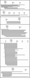
Schematic of all GRL antisense sequences isolated from exocytosis-deficient transformants. Sequences, which are represented as arrows, fall into six groups that correspond to the known GRLs. Nucleotide positions indicated at the top of each group are relative to the start codon of the indicated GRL. In cases where identical sequences were obtained from multiple clones arising from a single transformation, only a single arrow is shown. Six sequences (marked with an asterisk) were used to transform wild-type cells, and each of these antisense sequences conferred an exocytosis-deficient phenotype. All antisense sequences contain a portion of the 5′ untranslated region of their respective genes.
Figure 3.
Figure 3.
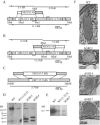
Disruption of GRL3, GRL4, and GRL7 through homologous recombination. (A) GRL3/4 genomic locus, showing the replacement by the NEO2 cassette of the first 508 aa of the GRL3 ORF. In A, B, and C, noncoding regions are shown in gray. (B) GRL3/4 locus, showing the replacement by the NEO2 cassette of the first 242 aa of the GRL4 ORF. (C) GRL7 genomic locus, showing replacement by the MTT-NEO cassette of the entire GRL7 ORF. (D) Southern blot of genomic DNA from GRL3 and GRL4 knockout lines, with probes generated from the cDNAs of the respective genes as described in Materials and Methods. An additional XbaI site in the NEO2 cassette results in hybridization of the probe to two smaller fragments; the remaining material at the size of the wild-type locus is transcriptionally silent micronuclear DNA, present 2N relative to the 45N of the transcriptionally active macronuclear DNA (a 22.5-fold decrease in signal, as shown). (E) Southern blot of a GRL7 knockout line, using a probe generated from the GRL7 cDNA. In this case, none of the GRL7 ORF remains for hybridization with the probe; the remaining signal is from detection of micronuclear DNA. (F) Electron micrographs of wild-type, ΔGRL3, ΔGRL4, and ΔGRL7 granules, showing the aberrant morphology and lack of a crystalline lattice in the knockout lines. Bars, 0.2, 0.25, 0.2, and 0.2 μm, respectively.
Figure 4.
Figure 4.
Affinity chromatography of proGrl1p complexes. (A) Identification of proGrl4p and Grl4p by Western blotting. Whole cell lysates of wild-type, ΔGRL4, and UC623 cells were resolved on a 17.5% gel and probed with the anti-Grl4p antibody on Western blots. Compared with ΔGRL4, the wild-type lane has two extra bands. The more abundant species, at 12 kDa, corresponds to the mature product generated from the amino-terminal half of proGrl4p (Verbsky and Turkewitz, 1998), whereas the lighter band at 47 kDa corresponds to proGrl4p. Consistent with these assignments, the latter is much more abundant, and the former absent, in a lysate from the UC623 mutant line that accumulates unprocessed proGrl proteins (Bowman et al., 2005). (B) proGrl4p and proGrl8p physically interact with proGrl1p. In wild-type cells, the endogenous GRL1 gene was replaced by a construct that adds a C-terminal 6His-tag to the GRL1 open reading frame and maintains expression from the endogenous promoter. Transformed cells were lysed as described in Materials and Methods, and proGrl1p-6His-containing complexes were purified from a high speed supernatant by affinity chromatography. The purified material (+His) was run on a 12.5% gel and probed with the anti-Grl1p, anti-Grl4p, and anti-Grl8p antibodies by Western blot. The control lanes (–) contain material purified from wild-type cells that were not transformed with the His-tag construct and contain material that was nonspecifically bound by the column. Specifically bound species, corresponding to Grl proproteins, are indicated by arrowheads. The electrophoretic positions of molecular weight standards are indicated on the right of each panel.
Figure 5.
Figure 5.
GRL4 is required for proGrl1p exit from the endoplasmic reticulum. (A) GRL4 is required for proGrl1p processing. Whole cell lysates of wild-type cells and the indicated ΔGRL strains were resolved on a 12.5% gel and probed with the anti-Grl1p antibody by Western blotting. The disruption of GRL3 or GRL8 has a partial inhibitory affect on the accumulation of processed Grl1p, whereas the disruption of GRL4 results in complete inhibition of proGrl1p processing. The ladder of intermediate bands in the ΔGRL3 and ΔGRL8 samples may be accounted for by weak cross-reactivity of the anti-Grl1p antibody for other Grl proproteins. The relative abundance of the proGrl and mature Grl species cannot be compared in this experiment, because the former seems to be more immunoreactive under Western blotting conditions (our unpublished data). (B) GRL1-GFP was expressed in wild-type cells and the indicated ΔGRL strains, and GFP localization was determined by confocal microscopy on live cells. Each confocal image shows the top surface of a single cell. In ΔGRL4 cells, the proGrl1p-GFP resides in a subplasma membrane reticulum. In all other strains, the fluorescent proteins localize to docked granules. Bars, 10 μm.
Figure 6.
Figure 6.
Overexpression of GRL1 results in the retention and crystallization of proGrl1p in the ER. (A) The accumulated proGrl1p in cadmium-induced cells constitutes a significant percentage of total cellular protein. Cells were transformed with the cadmium-inducible GRL1 expression vector. Whole cell lysates from either uninduced or 3.0 μg/ml cadmium-stimulated cells were analyzed by Coomassie Blue staining (12.5% polyacrylamide gel). A pronounced band at 60 kDa, corresponding to proGrl1p, is exclusively present in the induced sample (arrow). (B) ProGrl1p crystallizes in the ER. Cells overexpressing proGrl1p, induced as in A, were fixed and prepared for electron microscopy. Left, at low magnifications, a large number of straight-edged inclusions are visible. The asterisk indicates an inclusion that has formed in the envelope of endoplasmic reticulum that separates the nucleus (labeled N) from the cytosol. Bar, 1 μm. Right, high-magnification view reveals striated patterning in the inclusion bodies, indicating that the contents are crystalline. Docked ribosomes can be seen along the cytoplasmic membrane surface. Bar, 0.2 μm. (C) The extent of proGrl1p processing is independent of proGrl1p expression level. Expression of GRL1 from the inducible GRL1 expression vector was induced with the indicated cadmium concentrations for 6 h, and whole cell lysates of these cultures were resolved on a 12.5% gel and probed by Western blotting with anti-Grl1p antibody. The positions of proGrl1p and Grl1p are indicated. A cross-reactive band, marked by the asterisk, serves as a loading control.
Figure 7.
Figure 7.
Four new members of the GRL family. (A) Predicted structural features of Grl1p and of the four newly identified family members. (B) A phylogenetic tree displaying the evolutionary relationship between the Grlps. After aligning the protein sequences using the ClustalW algorithm, a phylogenetic tree was calculated using the maximum likelihood algorithm in the PHYLIP software suite. The branching pattern reveals two clusters of relatively closely-related paralogs (GRL1 and 2, and GRL5, 9, and 10) within a tree that is otherwise made up of deep lineages. (C) Alignment of the Grlps and a member of the Paramecium tmps, illustrating a tetrapeptide motif (boxed) that lies amino-terminal to the central basic stretch and to the carboxy-terminal basic stretch of Grl8p (shown in gray). With its substitution of a charged residue (Asp) within the motif, Grl2p deviates from the pattern established by the other family members.
Figure 8.
Figure 8.
Northern analysis of two novel GRLs. (A) Cellular RNA (9.4 μg) from growing (G) and starved (S) wild-type cells, labeled with a probe generated from the GRL1 (left blot) or GRL2 (right blot) open reading frames. At right on each blot is GRL1 (left blot) or GRL2 (right blot) RNA from in vitro transcription reactions, in loadings of 1 pg, 10 pg, 100 pg, and 1 ng. (B) For each of the blots in A, the 100-pg and 1-ng in vitro products were used to construct a linear scale. These scales were used to quantitate the amount of GRL1 and GRL2 RNA in G and S cells. (C) Northern blots as for A, probing for GRL7 and GRL6. (D) Analysis of the GRL7 and GRL6 blots as in B.
Figure 9.
Figure 9.
Localization of Grl2p and Grl6p to dense core granules by GFP chimeras. GRL2 and GRL6 were fused to GFP and expressed in growing cells. Shown for comparison are fusions of GFP with signal sequence alone (sig seq) or with a known granule content protein (GRL7). Bar, 10 μm.
Figure 10.
Figure 10.
Disruption of GRL2 and GRL6 through homologous recombination. (A) GRL2 genomic locus, showing the replacement by the MTT-NEO cassette of the entire GRL2 ORF. (B) Southern blot of a GRL2 knockout line, with a probe generated from the GRL2 cDNA. The remaining material at the size of the wild-type locus is transcriptionally silent micronuclear DNA. (C) GRL6 locus, showing the replacement by the MTT-NEO cassette of the entire GRL6 ORF. (D) Southern blot of a GRL6 knockout line, with a probe generated from the GRL6 cDNA; the remaining signal is from detection of micronuclear DNA. We used Southern blotting, with a probe generated from the NEO marker, to confirm that ΔGRL6 cells contained NEO at a single locus (our unpublished data).
Figure 11.
Figure 11.
Phenotypic analysis of GRL2 and GRL6 knockout lines. (A) Encapsulation assay. ΔGRL2 and ΔGRL6 cells were starved and treated with the dye Alcian Blue to induce capsule formation. Shown for comparison are wild-type and ΔGRL7 cells treated in the same manner. The dark circles in the ΔGRL7 cell are food vacuoles filled with ingested dye. Bar, 10 μm. (B) Electron micrographs of individual granules from ΔGRL2 and ΔGRL6 cells, showing that the contents are ordered. Bars, 0.2 μm.
Figure 12.
Figure 12.
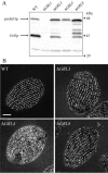
Granule synthesis defects in ΔGRL6 cells. (A) Granules in stationary phase cells were visualized using indirect immunofluorescence and mAb 4D11. Wild-type, ΔGRL2 and ΔGRL7 cells all show a dense pattern of docked DCGs. The DCGs in ΔGRL6 are comparatively sparse. Bar, 10 μm. (B) Images of live cells expressing Igr1p-GFP. Each image contains a cross-sectional view, showing the profiles of docked granules at the plasma membrane. The left side of the membrane faces the cytoplasm. Bar, 2 μm. (C) Immunodetection of Grl proteins (precursors and products) on Western blots of whole cell lysates. The bands corresponding to the precursor (arrow) and product (circle) of each Grl protein are indicated. ΔGRL6 cells accumulate a higher level of each precursor, and a lower level of each product, relative to wild type. It is important to note that these blots cannot be used to compare the relative levels of pro- and processed versions of each species, because some antibodies seem to be far more reactive on Western blots against the proprotein forms (Bowman and Turkewitz, unpublished).
Figure 13.
Figure 13.
Epistasis analysis of GRL6 function. (A) Southern blotting of whole cell DNA from wild type, ΔGRL6 in the CU428 (WT) background (same strain as in Figure 10), and two clones of MN173 ΔGRL6, using probes against GRT2 (loading control) and GRL6. Clone 7 has maintained GRL6 at wild-type levels, whereas clone 10 is equivalent to the established ΔGRL6 strain. The remaining signal is due to the micronuclear GRL6 copies, which are not expressed. (B) Flow cytometry of wild-type and ΔGRL6 cells to quantify Grl3p, a granule marker. Shown are two trials. In the first, wild-type (WT) cells are compared with ΔGRL6 cells, and the latter show a leftward shift representing a decrease in mean fluorescence per cell. In the second, wild-type cells are compared with MN173 clone 7 (wild-type level of GRL6) and clone 10 (ΔGRL6). Mean fluorescence in the two clones is similar. In both panels, the left-most curve represents cells with no 1° antibody. Identical results were obtained using mAb 4D11, which recognizes granule marker Grt2p. (C) Effect of GRL6 disruption on granule content. Shown is the ratio (mean, with SD) of FITC fluorescence in cells without GRL6 versus with GRL6. Mean FITC fluorescence was measured by flow cytometry, as in B, in cells labeled with mAbs 4D11 or 5E9. On the left, the ratio of fluorescence in ΔGRL6 cells versus wild type (n = 7). Disrupting GRL6 results in a decrease in mean fluorescence. On the right, the ratio of fluorescence in MN173ΔGRL6 versus MN173 (clone 10 vs. clone 7) (n = 5). In the MN173 background, disruption of GRL6 results in an increase in mean fluorescence.

Similar articles

Cited by

References

    1. Adoutte, A., Garreau de Loubresse, N., and Beisson, J. (1984). Proteolytic cleavage and maturation of the crystalline secretion products of Paramecium. J. Mol. Biol. 180, 1065–1081. - PubMed
    1. Andres, D. A., Rhodes, J. D., Meisel, R. L., and Dixon, J. E. (1991). Characterization of the carboxyl-terminal sequences responsible for protein retention in the endoplasmic reticulum. J. Biol. Chem. 266, 14277–14282. - PubMed
    1. Arvan, P., and Castle, D. (1998). Sorting and storage during secretory granule biogenesis: looking backward and looking forward. Biochem. J. 332, 593–610. - PMC - PubMed
    1. Arvan, P., and Castle, J. D. (1992). Protein sorting and secretion granule formation in regulated secretory cells. Trends Cell. Biol. 2, 327–331. - PubMed
    1. Arvan, P., Zhang, B. Y., Feng, L., Liu, M., and Kuliawat, R. (2002). Lumenal protein multimerization in the distal secretory pathway/secretory granules. Curr. Opin. Cell Biol. 14, 448–453. - PubMed

Publication types

MeSH terms

Substances

LinkOut - more resources