. 2005 Jun 15;25(24):5700-9.
doi: 10.1523/JNEUROSCI.0523-05.2005.

Bradykinesia induced by dopamine D2 receptor blockade is associated with reduced motor cortex activity in the rat

Affiliations

Bradykinesia induced by dopamine D2 receptor blockade is associated with reduced motor cortex activity in the rat

Louise C Parr-Brownlie et al. J Neurosci. .

Abstract

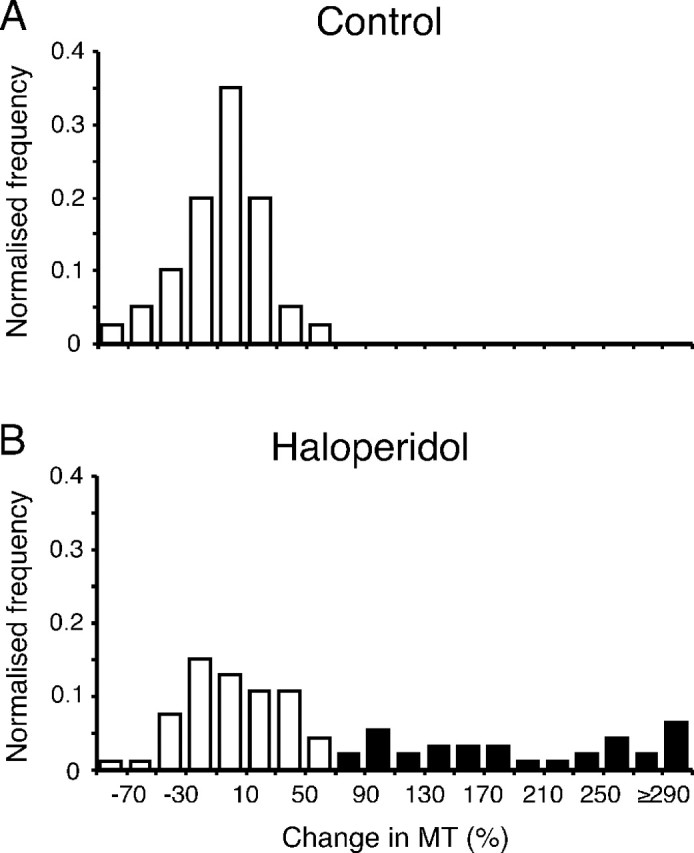
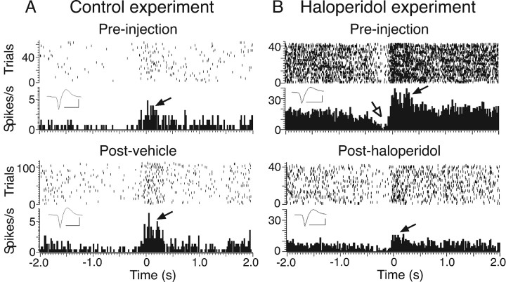
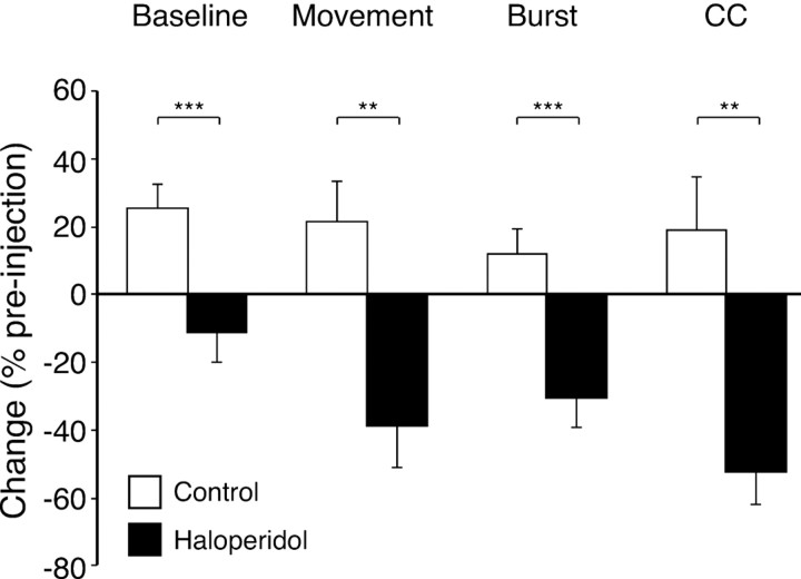
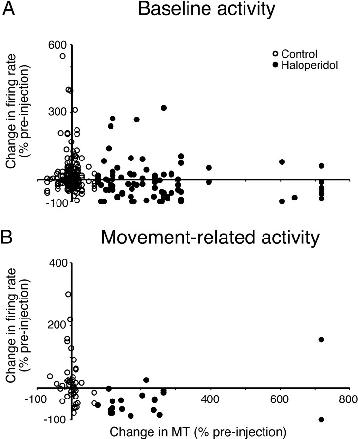
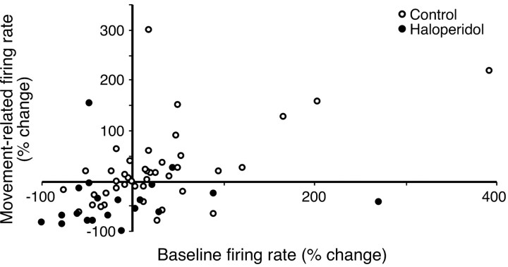
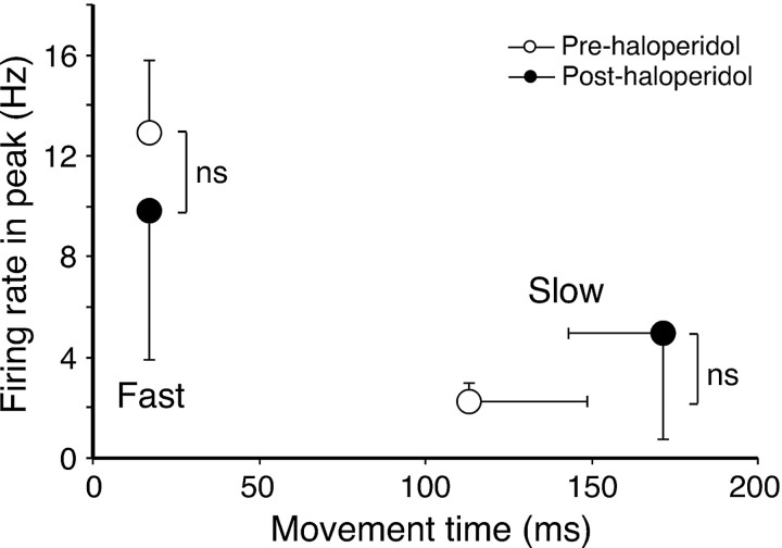
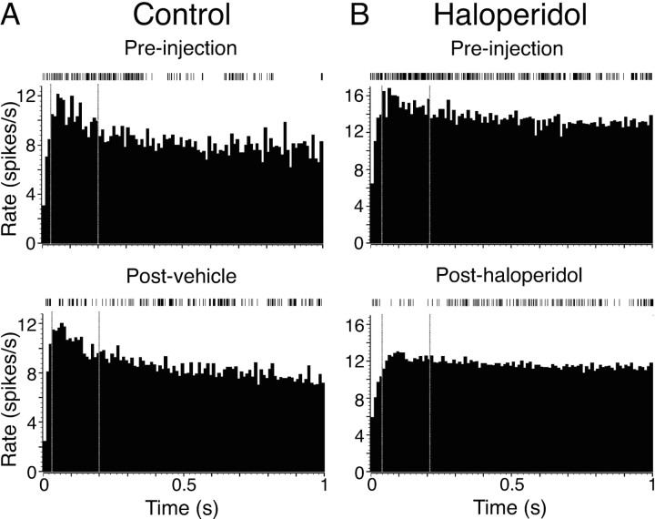
Disruption of motor cortex activity is hypothesized to play a major role in the slowed movement (bradykinesia) associated with reduced dopaminergic function. We recorded single neurons in the motor cortex of free-moving rats performing a forelimb-reaching task. The same neurons were examined before and after induction of bradykinesia with the D2 dopamine receptor antagonist haloperidol. Within-cell changes in the firing rate and firing pattern of individual cells and the correlation between simultaneously recorded cells after injection of haloperidol were statistically compared with vehicle-only control experiments. During haloperidol-induced bradykinesia (mean movement time increase, +231%), there was an average 11% decrease in baseline firing rate. Movement-related peaks in firing rate were more dramatically affected, with an overall reduction in peak amplitudes of 40%. Bradykinesia was also associated with decreased intensity of bursting and amplitude of cross-correlation peaks at rest. The results show for the first time that significant reductions can be detected in motor cortex activity at rest in animals with impaired ability to generate movements induced by reduced dopamine action and confirm that impaired movements are associated with reduced cortical activation. Together, these changes in neural activity may reduce recruitment and rate modulation of motor units in the spinal cord.


Figures

Figure 1.
Figure 1.
Identification of haloperidol-induced bradykinesia. Graphs show normalized frequency distributions of the change in movement time (MT) after vehicle (A) (control; n = 40) and haloperidol (B) (n = 93) injections. Bradykinetic experiments (filled columns) were defined as those exceeding the maximum percentage increase seen in control experiments.
Figure 2.
Figure 2.
Illustrative examples of dot raster and PETH analyses of baseline and movement-related cell activity and the effect of haloperidol. A, Cell recorded in a control experiment (injection of vehicle) with a significant movement-related excitation (filled arrow) and a nonsignificant inhibition. Top and bottom panels show preinjection and postinjection data, respectively. The dot rasters show each action potential in each trial, and the histograms show firing rate averaged across all trials (20 ms bins). Data are aligned to the termination of the extension phase of the reaching movement (time 0). Baseline activity was calculated from the first 1.5 s of the histogram. The inset shows averaged action potential waveforms for the spikes obtained in each recording. Calibration: 1 ms, 50 μV. B, A different cell recorded before and after induction of bradykinesia by haloperidol. Significant excitatory and inhibitory movement-related modulations in preinjection data are indicated by filled and open arrows, respectively.
Figure 3.
Figure 3.
Effect of haloperidol on measures of cell activity, expressed as a percentage change from preinjection values. Open bars show mean values from control (vehicle), and filled bars show changes with haloperidol-induced bradykinesia. Error bars represent SEM. Baseline, Baseline firing rate; Movement, movement-related firing rate; Burst, amplitude of autocorrelation peak; CC, amplitude of cross-correlation peak. **p ≤ 0.01, ***p ≤ 0.001; one-factor ANOVAs or Mann-Whitney U tests, comparing vehicle and haloperidol experiments.
Figure 4.
Figure 4.
Relationship between change in movement time and cell activity measures. A, Scatter plot of percentage change in baseline firing rate and percentage change in movement time (MT) for cells recorded in vehicle injection control (open circles) or bradykinetic haloperidol experiments (filled circles). B, Scatter plot of percentage change in movement time and percentage change in excitatory movement-related firing rate.
Figure 5.
Figure 5.
Relationship between change in movement-related firing rate and change in baseline firing rate. Data are shown for each recorded cell in vehicle injection control experiments (open circles) or bradykinetic haloperidol experiments (filled circles).
Figure 6.
Figure 6.
Within-session analysis of movement-related firing rates. The graph shows mean values for movement-related firing rates calculated for selected slow and fast trials obtained from preinjection (open circles) and posthaloperidol (filled circles) periods. Error bars represent SEM. ns, Nonsignificant difference; paired t test.
Figure 7.
Figure 7.
Illustrative examples of changes in burst amplitude during quiet rest. Autocorrelograms show average firing rate for 1 s after each spike. A, Cell recorded in prevehicle and postvehicle periods of a control experiment. B, A different cell recorded before and after haloperidol injection. Vertical dotted lines indicate the region from which the mean amplitude of peaks was determined (region >2 SDs of histogram baseline mean, determined from preinjection histogram). Above each autocorrelogram is a 20 s sample of spike train during the rest period.

References

    1. Albin RL, Young AB, Penney JB (1989) The functional anatomy of basal ganglia disorders. Trends Neurosci 12: 366-375. - PubMed
    1. Alexander GE, Crutcher MD (1990) Functional architecture of basal ganglia circuits: neural substrates of parallel processing. Trends Neurosci 13: 266-271. - PubMed
    1. Berardelli A, Rothwell JC, Thompson PD, Hallett M (2001) Pathophysiology of bradykinesia in Parkinson's disease. Brain 124: 2131-2146. - PubMed
    1. Bergman H, Wichmann T, Karmon B, DeLong MR (1994) The primate subthalamic nucleus. II. Neuronal activity in the MPTP model of parkinsonism. J Neurophysiol 72: 507-520. - PubMed
    1. Bonfiglioli C, DeBerti G, Nichelli P, Nicoletti R, Castiello U (1998) Kinematic analysis of the reach to grasp movement in Parkinson's and Huntington's disease subjects. Neuropsychologia 36: 1203-1208. - PubMed

Publication types

LinkOut - more resources