. 2005 Jun 15;25(24):5710-9.
doi: 10.1523/JNEUROSCI.0274-05.2005.

Conditional rhythmicity of ventral spinal interneurons defined by expression of the Hb9 homeodomain protein

Affiliations

Conditional rhythmicity of ventral spinal interneurons defined by expression of the Hb9 homeodomain protein

Jennifer M Wilson et al. J Neurosci. .

Abstract

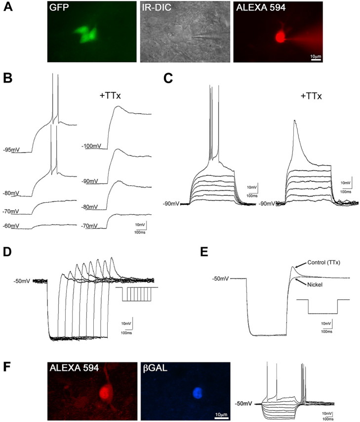
The properties of mammalian spinal interneurons that underlie rhythmic locomotor networks remain poorly described. Using postnatal transgenic mice in which expression of green fluorescent protein is driven by the promoter for the homeodomain transcription factor Hb9, as well as Hb9-lacZ knock-in mice, we describe a novel population of glutamatergic interneurons located adjacent to the ventral commissure from cervical to midlumbar spinal cord levels. Hb9+ interneurons exhibit strong postinhibitory rebound and demonstrate pronounced membrane potential oscillations in response to chemical stimuli that induce locomotor activity. These data provide a molecular and physiological delineation of a small population of ventral spinal interneurons that exhibit homogeneous electrophysiological features, the properties of which suggest that they are candidate locomotor rhythm-generating interneurons.


Figures

Figure 1.
Figure 1.
GFP+ motoneurons and interneurons in the spinal cord of the postnatal Hb9:GFP mouse. A, Fluorescent micrographs of 30 μm upper lumbar spinal cord sections illustrate GFP-expressing neurons in the ventral spinal cord (Ai), which include somatic and sympathetic motoneurons, identified by colocalization with Fluorogold (Aii). Note the labeling of sympathetic preganglionic neurons in the intermediolateral cell column and central autonomic area, dorsal to the central canal. B-D, High-magnification images corresponding to marked regions in A illustrate somatic (B) and sympathetic preganglionic (C) motoneuron GFP expression, identified by Fluorogold labeling. D, GFP+ neurons that abut the ventral commissure are not Fluorogold positive and are therefore interneurons.
Figure 2.
Figure 2.
Medial lamina VIII interneurons are Hb9+. A, In Hb9:GFP × Hb9nlsLacZ/+ offspring, low-power confocal projections show the distributions of GFP, β-gal, and ChAT+ neurons in the spinal cord. B, Higher-magnification images indicate that medial lamina VIII interneurons are β-gal+ (and thus Hb9+) but ChAT-. The midline is to the left of these cells. A separate, small population of interneurons in ventral medial lamina VIII coexpressed GFP with ChAT, but these neurons lacked β-gal expression (Hb9) (data not shown). C, Most GFP+ scattered interneurons throughout the ventral horn are both β-gal- and ChAT-. D, Motoneurons identified by ChAT immunoreactivity are β-gal+. Note that not all motoneurons are GFP+. Scale bars: B-D, 20 μm.
Figure 3.
Figure 3.
Hb9+ interneurons are glutamatergic and do not appear to provide a major input to motoneurons. A, Confocal images of fluorescence in situ hybridization studies reveal expression of VGLUT2 mRNA (Ai) in cells that express GFP (Aii). All medial GFP+ neurons contain mRNA for VGLUT2 (Aiv-Avi), including the clustered neurons abutting the ventral commissure recognized as Hb9+ (Av is an enlargement of the box in Aiv), as well as populations of neurons located more laterally (Avi). Scale bar: (in Aii) for Ai-Aiv, 100 μm; (in Avi) for Av-Avi, 50 μm. Avii, Neurolucida schematic drawings illustrate the corresponding clustering of Hb9+ interneurons abutting the ventral commissure in the Hb9:GFP × Hb9nlsLacZ/+ double-transgenic mice. Note that the GFP+ cells that express VGLUT2 (Aiii) include the Hb9+ interneurons (Avii). B, GFP+ motoneuron somata and proximal dendrites in the lateral motor column in L2 are devoid of VGLUT2+ GFP+ terminals. This image is representative of the thorough scanning of three 70 μm sections from L2 in each of three mice. The single VGLUT2+ GFP+ terminal in this frame is marked with an arrow and magnified in the inset. Scale bar, 10 μm.
Figure 4.
Figure 4.
Excitatory and inhibitory inputs to Hb9+ interneurons. A-D, Single optical section confocal micrographs illustrate VGLUT1+ (A) and VGLUT2+ (B) axon terminals apposed to medial lamina VIII GFP+ neurons. Note that some VGLUT2+ terminals apposed to the Hb9+ interneurons are also GFP+ and may therefore reflect self- or mutual excitation. There are also GAD-67+ (C) and GlyT2+ (D) terminals apposed to the Hb9+ interneurons. Scale bar, 10 μm. E, In the Hb9:GFP × Hb9nlsLacZ/+ double-transgenic mouse, 5-HT+ terminals make contact with GFP+ interneurons that are also β-gal+ (Hb9+). Scale bar, 1 μm.
Figure 5.
Figure 5.
The electrophysiological profile of Hb9+ interneurons. A, Interneurons identified by the presence of GFP were patch clamped using IR-differential interference contrast (DIC) and filled with Alexa 594. B, The PIR that evokes action potential firing is dependent on the preceding voltage. In TTX, the amplitude of the PIR is dependent on previous membrane voltage. Hyperpolarizing pulses of 500 ms duration were elicited from -50 mV. C, Injection of linearly increasing current pulses from a holding potential of -90 mV revealed an all-or-none depolarization that, after reaching threshold, led to a burst of action potentials. In the presence of TTX (1 μm), an underlying low-amplitude spike was revealed. D, The amplitude of the PIR (in TTX) is dependent on the duration of preceding hyperpolarization. E, The PIR is blocked with nickel (100 μm). F, In Hb9:GFP × Hb9nlsLacZ/+ double-transgenic mouse slices, recorded cell somata were filled with the intracellular dye Alexa 594. Immunohistochemical processing after recording revealed colocalization of Alexa 594 with β-gal, demonstrating that these cells (type 1) are indeed Hb9+.
Figure 6.
Figure 6.
Larger lamina VIII interneurons (type 2) exhibit a time-dependent sag, which is typical of that mediated by Ih, and are electrotonically coupled. A, Interneurons identified by the presence of GFP were patch clamped under IR-differential interference contrast (DIC) optics and filled with Alexa 594. B, Membrane-voltage responses to increasing injections of current pulses reveal a characteristic sag in membrane voltage at potentials greater than -80 mV. In addition, a small “hump” is evident on the break of the hyperpolarizing current pulse that could reach threshold for single action potential generation. C, These interneurons were spontaneously active at resting potentials with voltage-independent, biphasic subthreshold oscillations in membrane potential evident after hyperpolarization of the membrane (arrows). This indicates that these cells were likely electrotonically coupled to active neurons.
Figure 7.
Figure 7.
Hb9+ interneurons are active during locomotion and are conditionally rhythmic. A, Adult mice were subjected to 90 min of walking to induce activity-dependent Fos expression. Confocal micrographs illustrate Fos immunoreactivity in the clustered medial lamina VIII Hb9+ interneurons (arrows) but not in the more lateral larger GFP+ (Hb9-) interneurons (asterisks). In the control condition, mice slept before perfusion, and the Hb9+ interneurons were Fos-. Experimental and control experiments were both conducted at mid-day. Low-magnification insets indicate location of the interneurons abutting the ventral commissure. B-D, Application of the neurotransmitters 5-HT (20 μm), dopamine (DA; 50 μm), and NMDA (20 μm) in the presence of TTX (1 μm) induced two distinct waveforms of endogenous activity in Hb9+ cells. B, In some Hb9+ neurons, small-amplitude subthreshold oscillations were evoked. The amplitude of these oscillations was voltage independent. C, In other Hb9+ neurons, large oscillations in membrane potential could reach threshold for calcium spikes. Note the all-or-none nature of the activation of the calcium spike. The amplitude of these oscillations was voltage dependent, with a voltage window where calcium spikes were evoked. This large calcium spike component of the oscillations was blocked by nickel (100 μm), leaving small-amplitude oscillations. D, The frequency of the large-amplitude oscillations was voltage dependent, whereas the frequency of the smaller-amplitude oscillations was independent of membrane potential.

Similar articles

Cited by

References

    1. Aizenman CD, Linden DJ (1999) Regulation of the rebound depolarization and spontaneous firing patterns of deep nuclear neurons in slices of rat cerebellum. J Neurophysiol 82: 1697-1709. - PubMed
    1. Arber S, Han B, Mendelsohn M, Smith M, Jessell TM, Sockanathan S (1999) Requirement for the homeobox gene Hb9 in the consolidation of motor neuron identity. Neuron 23: 659-674. - PubMed
    1. Arshavsky YI, Deliagina TG, Orlovsky GN, Panchin YV, Popova LB, Sadreyev RI (1998) Analysis of the central pattern generator for swimming in the mollusk Clione Ann NY Acad Sci 860: 51-69. - PubMed
    1. Baev KV, Degtiarenko AM, Zavadskaia TV, Kostiuk PG (1979) Impulse activity of interneurons of the lumbar portion of the spinal cord during the late long discharges in the motor nerves of immobilized thalamic cats. Neirofiziologiia 11: 236-244. - PubMed
    1. Bannatyne BA, Edgley SA, Hammar I, Jankowska E, Maxwell DJ (2003) Networks of inhibitory and excitatory commissural interneurons mediating crossed reticulospinal actions. Eur J Neurosci 18: 2273-2284. - PMC - PubMed

Publication types