. 2005 Jun 16;435(7044):969-73.
doi: 10.1038/nature03703.

In vivo imaging of specialized bone marrow endothelial microdomains for tumour engraftment

Affiliations

In vivo imaging of specialized bone marrow endothelial microdomains for tumour engraftment

Dorothy A Sipkins et al. Nature. .

Abstract

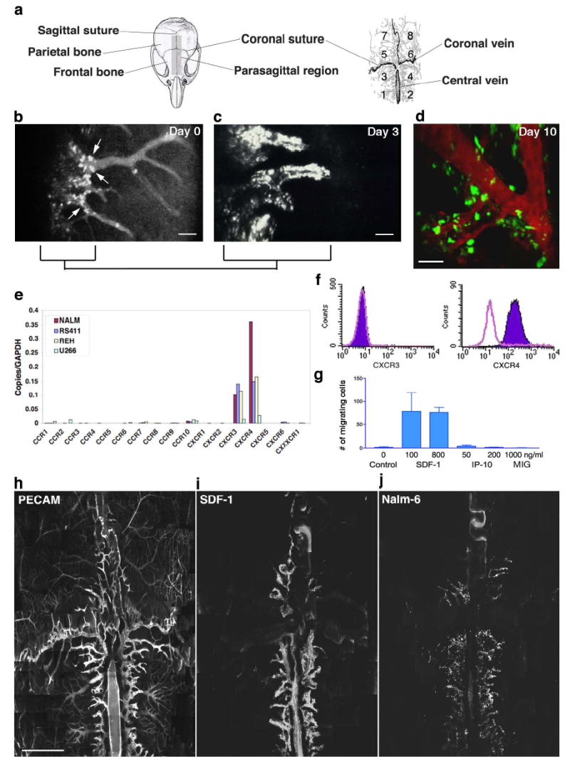
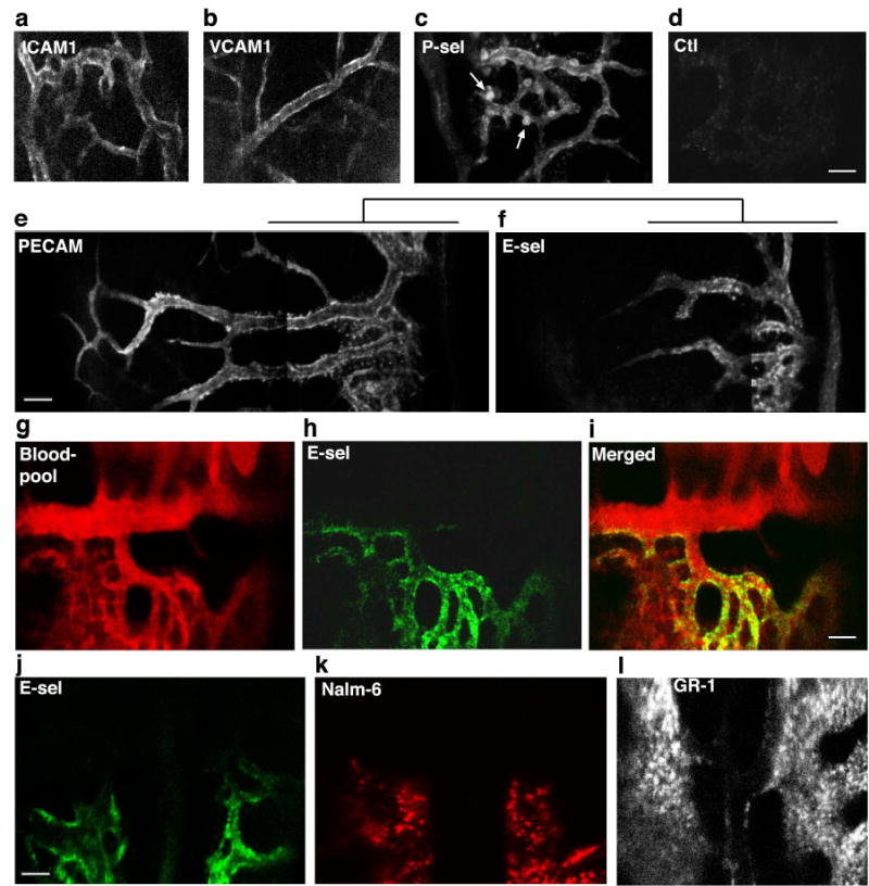
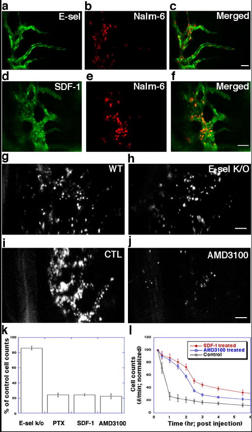
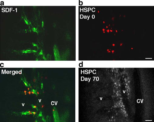
The organization of cellular niches is known to have a key role in regulating normal stem cell differentiation and regeneration, but relatively little is known about the architecture of microenvironments that support malignant metastasis. Using dynamic in vivo confocal imaging, here we show that murine bone marrow contains unique anatomic regions defined by specialized endothelium. This vasculature expresses the adhesion molecule E-selectin and the chemoattractant stromal-cell-derived factor 1 (SDF-1) in discrete, discontinuous areas that influence the homing of a variety of tumour cell lines. Disruption of the interactions between SDF-1 and its receptor CXCR4 inhibits the homing of Nalm-6 cells (an acute lymphoblastic leukaemia cell line) to these vessels. Further studies revealed that circulating leukaemic cells can engraft around these vessels, suggesting that this molecularly distinct vasculature demarcates a microenvironment for early metastatic tumour spread in bone marrow. Finally, purified haematopoietic stem/progenitor cells and lymphocytes also localize to the same microdomains, indicating that this vasculature might also function in benign states to demarcate specific portals for the entry of cells into the marrow space. Specialized vascular structures therefore appear to delineate a microenvironment with unique physiology that can be exploited by circulating malignant cells.


Conflict of interest statement

Competing interests statement The authors declare that they have no competing financial interests.

Figures

Figure 1
Figure 1
Leukemic cell homing/engraftment in mouse skull BM in vivo. a. Cartoons of the mouse skull and vascular anatomy, divided into quadrants for reference. b. Nalm-6 cells bound to parasagittal vascular segments (arrows) 1h post-injection (area 4). Scale bar = 200 μm. c–d. Peri-vascular cell growth 3 and 10 days post-injection. Scale bars = 100 μm. e. Quantitative RT-PCR of Nalm-6 and other tumor cell lines reveals abundant mRNA for CXCR4 and CXCR3. f. Flow cytometry of Nalm-6 demonstrates surface expression of CXCR4 only. g. In vitro chemotaxis of Nalm-6 toward the CXCR ligand SDF-1 but not toward the CXCR3 ligands IP-10 or MIG. Error bars represent standard error of the mean. h. BM vasculature labelled with anti-CD31 (assembled from approximately 500 images, scale bar = 1 mm). Signal dampening in lateral regions is due to variations in tissue optical properties (Supplementary Fig. 10). i. Montage image assembled from a separate mouse showing restricted SDF-1 expression (see also Supplementary Fig. 3, PECAM-1/SDF-1 co-label). j. Nalm-6 homing to SDF-1-expressing regions in this montage image from a third mouse.
Figure 2
Figure 2
In vivo immunofluorescence microscopy of vascular CAM expression in BM. a–c. Representative images of ICAM-1 (area 6), VCAM-1 (area 8) and P-selectin (area 4). Each are expressed throughout the BM vasculature. Arrows indicate P-selectin+ megakaryocytes. d. Isotype control. e–f. Montage images of extended PECAM-1 expression vs. restricted E-selectin expression (both area 3). g–i. In this image series from area 4/6, the BM blood pool (red) is delineated by a non-specific fluorescent antibody signal. E-selectin expression (green) is restricted to one wall of a large collecting vein and to sinusoidal microvasculature in area 4. j–l. Correspondence of E-selectin expression (green) with Nalm-6 (red) homing patterns. This is contrasted with anti-GR1 (myeloid differentiation antigen) staining of the same region (midline, intersection of areas 3–6). Scale bars = 100 μm.
Figure 3
Figure 3
Leukemic cell homing to SDF-1+E-selectin+ microvascular domains is inhibited by SDF-1/CXCR4 blockade. a–c. Co-localization of E-selectin expression (green) and Nalm-6 cell homing (red, 30 minutes post-injection). (parasagittal region of area 4). d–f. Co-localization of SDF-1 vascular expression (green) and Nalm-6 cell homing (red, 30 minutes post-injection) (area 3). g–h. Cell homing is slightly decreased (<20%) in the E-selectin knockout mouse compared to wild-type control (1h post-injection). i–j. Nalm-6 homing is inhibited by AMD3100-CXCR4 blockade. k. Quantitation of effect of SDF-1/CXCR4 inhibition on cell homing to BM microdomains by pertussis toxin (PTX), SDF-1-desensitization and AMD3100. Error bars represent standard error of the mean. l. Enumeration of circulating leukemic cells by in vivo flow cytometry demonstrates that Nalm-6 blocked from BM homing by SDF-1-desensitization or single-dose AMD3100 remain in peripheral circulation. As the treatment effects wear off, cells correspondingly exit the circulation. Scale bars = 100 μm. Error bars represent standard error of the mean.
Figure 4
Figure 4
HSPC homing to SDF-1-positive vascular microdomains. a–c. Co-localization of SDF-1 (green) and HSPC (red) 2h post-injection (parasagittal region of area 3). d. The same mouse imaged 70 days post-HSPC injection (approximately the same area as in a–c). Although fluorescence signal intensity is diminished due to HSPC proliferation, engrafted cells are apparent in the perivascular spaces. Scale bars = 100 μm. CV=central vein, v=venule.

Similar articles

Cited by

References

    1. Fuchs E, Tumbar T, Guasch G. Socializing with the neighbors: Stem cells and their niche. Cell. 2004;116:769–778. - PubMed
    1. Chambers AF, Groom AC, MacDonald IC. Dissemination and growth of cancer cells in metastatic sites. Nat Rev Cancer. 2002;2:563–72. - PubMed
    1. Honn KV, Tang DG. Adhesion molecules and tumor cell interaction with endothelium and endothelial matrix. Cancer and Metastasis Reviews. 1992;11:353–375. - PubMed
    1. Burger JA, Kipps TJ. Chemokine receptors and stromal cells in the homing and homeostasis of chronic lymphocytic leukemia B cells. Leukemia & Lymphoma. 2002;43:461–466. - PubMed
    1. Reuss-Borst MA, Klein G, Waller HD, Muller CA. Differential expression of adhesion molecules in acute leukemia. Leukemia. 1995;9:869–874. - PubMed

Publication types