

Immune responses to p53 in patients with cancer: enrichment in tetramer+ p53 peptide-specific T cells and regulatory T cells at tumor sites
- PMID: 15959774
- PMCID: PMC11032902
- DOI: 10.1007/s00262-005-0670-9
Immune responses to p53 in patients with cancer: enrichment in tetramer+ p53 peptide-specific T cells and regulatory T cells at tumor sites
Abstract
Objective: A majority of human cancers, including head and neck cancer (HNC), "overexpress" p53. Although T cells specific for wild-type (wt) sequence p53 peptides are detectable in the peripheral blood of patients with HNC, it is unknown whether such T cells accumulate in tumor-involved tissues. Also, the localization of "regulatory" T cells (Treg) to tumor sites in HNC has not been investigated to date.
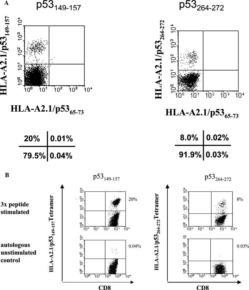
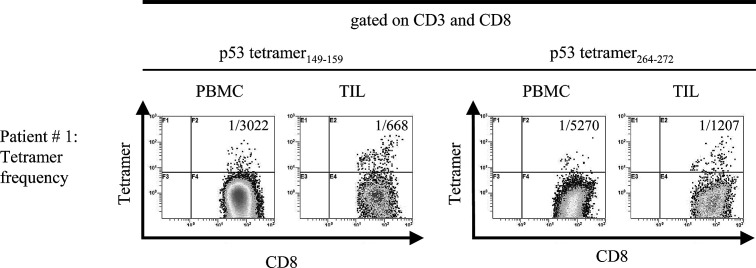
Methods: Tumor infiltrating lymphocytes (TIL), tumor-involved or non-involved lymph node lymphocytes (LNL) and peripheral blood mononuclear cells (PBMC) were obtained from 24 HLA-A2.1+ patients with HNC. Using tetramers and four-color flow cytometry, the frequency of Treg and CD3+CD8+ T cells specific for wt p53 epitopes as well as their functional attributes were determined.
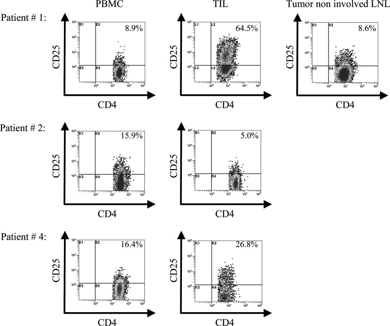
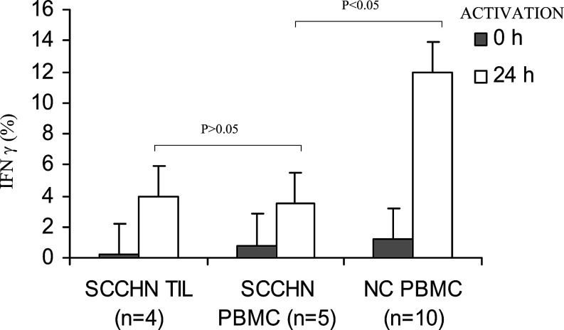
Results: The CD3+CD8+ tetramer+ cell frequency was significantly higher (P<0.001) in TIL than autologous PBMC as was the percentage of CD4+CD25+ T cells (P<0.003). TIL were enriched in FOXp3+, GITR+ and CTLA-4+ Treg. CD8+ TIL had low Zeta expression and produced little IFN-gamma after ex vivo stimulation relative to autologous PBMC or PBMC from NC.
Conclusions: Anti-wt p53 epitope-specific T cells and Treg preferentially localize to tumor sites in patients with HNC. However, despite enrichment in tumor peptide-specific T cells, the effector cell population (CD3+CD8+) in TIL or PBMC was unresponsive to activation in the tumor microenvironment enriched in Treg.
Figures
References
-
- Chikamatsu K, Nakano K, Storkus WJ, Appell E, Lotze MT, Whiteside TL, DeLeo AB. Generation of anti-p53 cytotoxic T lymphocytes from human peripheral blood using autologous dendritic cells. Clin Cancer Res. 1999;5:1281. - PubMed
-
- Elder EM, Whiteside TL (1992) Processing of tumors for vaccine and/or tumor infiltrating lymphocytes. In: Friedman H, Rose NR, deMacario EC, Fahey JL, Friedman H, Penn GM (Eds) Manual of clinical laboratory immunology, vol 123, 4th edn. American Society of Microbiology, Washington, p 817
-
- Gnjatic S, Cai Z, Viguier M, Chouaib S, Guillet J-G, Choppin J. Accumulation of the p53 protein allows recognition by human CTL of a wild-type p53 epitope presented by breast carcinomas and melanomas. J Immunol. 1998;160:328. - PubMed
-
- Harris CC. Structure and function of the p53 tumor suppressor gene: clues and rational cancer therapeutic strategies. J Natl Cancer Inst. 1996;88:1442. - PubMed
Publication types
MeSH terms
Substances
Grants and funding
LinkOut - more resources
Full Text Sources
Medical
Research Materials
Miscellaneous
