. 2005 Jun;16(6):741-51.
doi: 10.1089/hum.2005.16.741.

Stability of lentiviral vector-mediated transgene expression in the brain in the presence of systemic antivector immune responses

Affiliations

Stability of lentiviral vector-mediated transgene expression in the brain in the presence of systemic antivector immune responses

Evelyn Abordo-Adesida et al. Hum Gene Ther. 2005 Jun.

Abstract

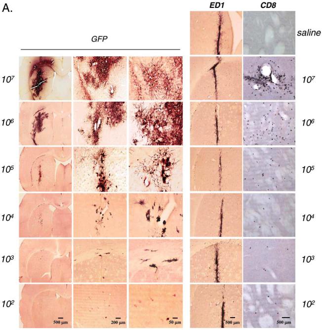
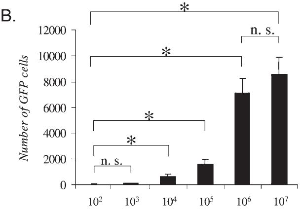
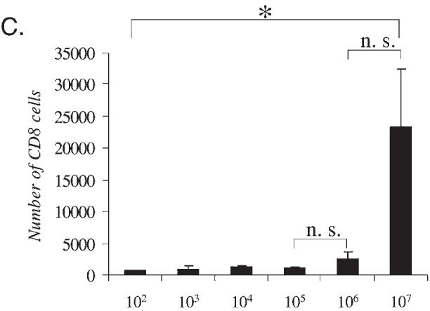
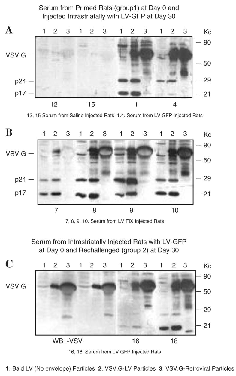
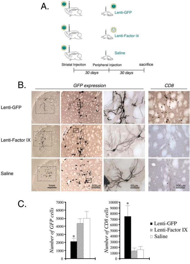
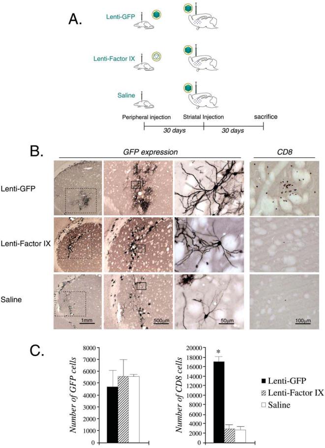
Lentiviral vectors are promising tools for gene therapy in the CNS. It is therefore important to characterize their interactions with the immune system in the CNS. This work characterizes transgene expression and brain inflammation in the presence or absence of immune responses generated after systemic immunization with lentiviral vectors. We characterized transduction with SIN-LV vectors in the CNS. A dose-response curve using SIN-LV-GFP demonstrated detectable transgene expression in the striatum at a dose of 10(2), and maximum expression at 10(6), transducing units of lentiviral vector, with minimal increase in inflammatory markers between the lowest and highest dose of vector injected. Our studies demonstrate that injection of a lentiviral vector into the CNS did not cause a measurable inflammatory response. Systemic immunization after CNS injection, with the lentiviral vector expressing the same transgene as a vector injected into the CNS, caused a decrease in transgene expression in the CNS, concomitantly with an infiltration of inflammatory cells into the CNS parenchyma at the injection site. However, peripheral immunization with a lentiviral vector carrying a different transgene did not diminish transgene expression, or cause CNS inflammation. Systemic immunization preceding injection of lentiviral vectors into the CNS determined that preexisting antilentiviral immunity, regardless of the transgene, did not affect transgene expression. Furthermore, we showed that the transgene, but not the virion or vector components, is responsible for providing antigenic epitopes to the activated immune system, on systemic immunization with lentivirus. Low immunogenicity and prolonged transgene expression in the presence of preexisting lentiviral immunity are encouraging data for the future use of lentiviral vectors in CNS gene therapy. In summary, the lentiviral vectors tested induced undetectable activation of innate immune responses, and stimulation of adaptive immune responses against lentiviral vectors was effective in causing a decrease in transgene expression only if the immune response was directed against the transgene. A systemic immune response against vector components alone did not cause brain inflammation, possibly because vector-derived epitopes were not being presented in the CNS.


Figures

FIG. 1
FIG. 1
Dose response for Lenti-GFP in the CNS: Transgene expression and markers for innate inflammation. Immunohistochemistry for GFP, ED1, and CD8 was performed in serial brain sections of rats injected with Lenti-GFP via the striatum and perfused—fixed 30 days later. (A) Increase in expression of the transgene GFP from 102 to 107 TU injected directly into the striatum. The innate inflammatory response is illustrated by the influx of macrophage/microglial cells (immunoreactive for ED1) and T cells (immunoreactive for CD8). The number of immunoreactive GFP+ cells increased with injection of 102 to 106 TU; there was no difference at either dose in the amount of cells immunoreactive for ED1. However, the number of CD8+ cells was significantly increased at 107 TU. Quantification of these data is shown in (B) and (C), which illustrates that there is no further increase in transduced cells after the injection of 107 IU of Lenti-GFP into the striatum (B) and that there is a significant increase in CD8+ cells with injection of 107 IU (C). Scale bars are shown in (A). *p < 0.05.
FIG. 1
FIG. 1
Dose response for Lenti-GFP in the CNS: Transgene expression and markers for innate inflammation. Immunohistochemistry for GFP, ED1, and CD8 was performed in serial brain sections of rats injected with Lenti-GFP via the striatum and perfused—fixed 30 days later. (A) Increase in expression of the transgene GFP from 102 to 107 TU injected directly into the striatum. The innate inflammatory response is illustrated by the influx of macrophage/microglial cells (immunoreactive for ED1) and T cells (immunoreactive for CD8). The number of immunoreactive GFP+ cells increased with injection of 102 to 106 TU; there was no difference at either dose in the amount of cells immunoreactive for ED1. However, the number of CD8+ cells was significantly increased at 107 TU. Quantification of these data is shown in (B) and (C), which illustrates that there is no further increase in transduced cells after the injection of 107 IU of Lenti-GFP into the striatum (B) and that there is a significant increase in CD8+ cells with injection of 107 IU (C). Scale bars are shown in (A). *p < 0.05.
FIG. 2
FIG. 2
Immune response to VSV-pseudotyped lentiviral vector particles. (A and B) Sera from rats injected subcutaneously with saline (rats 12 and 15), Lenti-GFP (rats 1 and 4), or Lenti-FIX (rats 7–10), and 30 days later injected via the brain with Lenti-GFP, were tested after another 30 days for the presence of antibodies against vector particles by Western blot. The following types of particles were loaded on the gel: lane 1, bald lentiviral particles (no VSV); lane 2, VSV-G-pseudotyped lentiviral particles; lane 3, VSV-G-pseudotyped retroviral particles. The expected migration of HIV-1 p24 capsid, p17 matrix protein, and VSV-G envelope protein is indicated. (C) Left: Anti-VSV antibodies were also used to identify VSV-G protein in the blot. Right: Two representative blots of sera from rats (rats 16 and 18) first injected via the brain with Lenti-GFP, challenged subcutaneously after 30 days with Lenti-GFP and Lenti-FIX, and analyzed after another 30 days.
FIG. 3
FIG. 3
CNS immune responses to lentiviral vector: Systemic immunization after CNS injection of lentiviral vector. (A) Schematic illustration of the experimental procedure, in which rats were injected with 105 TU of Lenti-GFP intrastriatally and immunized systemically 30 days later with either Lenti-GFP, Lenti-FIX, or saline. (B) GFP expression, and T cell (CD8+) infiltration, in the striatum of each group of animals 30 days after systemic immunization. (C) Quantification of the serial sections in (B), and statistical analysis (*p < 0.05; versus Lenti-FIX and saline groups). In (B), of the columns labeled GFP, the leftmost column illustrates a low-power view of the striatum, and the middle and right columns illustrate increasingly higher power views of striatal cells expressing GFP. Scale bars are shown in (B).
FIG. 4
FIG. 4
CNS immune responses to lentiviral vector: Systemic immunization preceding CNS injection of lentiviral vector. (A) Schematic illustration of the experimental procedure, in which rats were injected with Lenti-GFP, Lenti-FIX, or saline systemically, and injected intrastriatally 30 days later with 105 TU of Lenti-GFP. (B) GFP expression, and T cell (CD8+) infiltration, in the striatum of each group of animals. (C) Quantification of the serial sections in (B), and statistical analysis (*p < 0.05; versus Lenti-FIX and saline groups). In (B), of the columns labeled GFP, the leftmost column illustrates a low-power view of the striatum, and the middle and right columns illustrate increasingly higher power views of striatal cells expressing GFP. Scale bars are shown in (B).

References

    1. AZZOUZ M, RALPH GS, STORKEBAUM E, WALMSLEY LE, MITROPHANOUS KA, KINGSMAN SM, CARMELIET P, MAZARAKIS ND. VEGF delivery with retrogradely transported lentivector prolongs survival in a mouse ALS model. Nature. 2004;429:413–417. - PubMed
    1. BENNETT J. Immune response following intraocular delivery of recombinant viral vectors. Gene Ther. 2003;10:977–982. - PubMed
    1. BIFFI A, DE PALMA M, QUATTRINI A, DEL CARRO U, AMADIO S, VISIGALLI I, SESSA M, FASANO S, BRAMBILLA R, MARCHESINI S, BORDIGNON C, NALDINI L. Correction of metachromatic leukodystrophy in the mouse model by transplantation of genetically modified hematopoietic stem cells. J. Clin. Invest. 2004;113:1118–1129. - PMC - PubMed
    1. BLOMER U, NALDINI L, KAFRI T, TRONO D, VERMA IM, GAGE FH. Highly efficient and sustained gene transfer in adult neurons with a lentivirus vector. J. Virol. 1997;71:6641–6649. - PMC - PubMed
    1. CONSIGLIO A, QUATTRINI A, MARTINO S, BENSADOUN JC, DOLCETTA D, TROJANI A, BENAGLIA G, MARCHESINI S, CESTARI V, OLIVERIO A, BORDIGNON C, NALDINI L. In vivo gene therapy of metachromatic leukodystrophy by lentiviral vectors: Correction of neuropathology and protection against learning impairments in affected mice. Nat. Med. 2001;7:310–316. - PubMed

Publication types

Substances

LinkOut - more resources