

Morphometric and immunohistochemical study of the omasum of red deer during prenatal development
- PMID: 15960765
- PMCID: PMC1571522
- DOI: 10.1111/j.1469-7580.2005.00409.x
Morphometric and immunohistochemical study of the omasum of red deer during prenatal development
Abstract
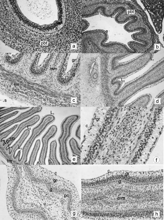
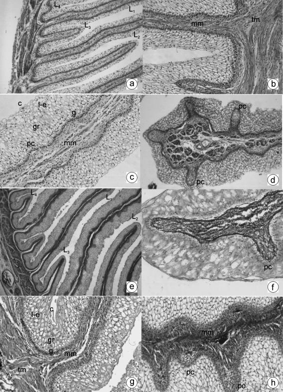
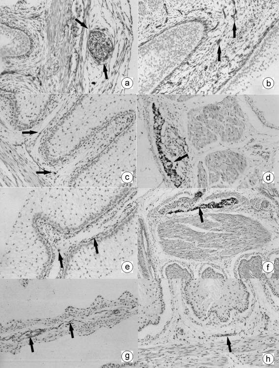
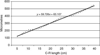
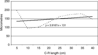
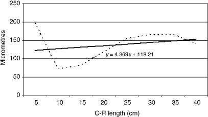
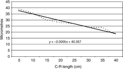
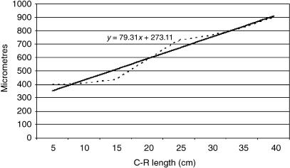
The red deer is an important study species because of its value in the national economy and because it provides a wealth of genetic material. To date, there has been little research into the prenatal development of the stomach of ruminants, and none of the red deer. We therefore performed a histological evaluation of the ontogenesis of the omasum in the red deer. Histomorphometric and immunohistochemical analyses were carried out on 50 embryos and fetuses of deer from the initial stages of prenatal life until birth. For test purposes, the animals were divided into five experimental groups: Group I (1.4-3.6 cm crown-rump length, CRL; 30-60 days, 1-25% of gestation); Group II (4.5-7.2 cm CRL; 67-90 days, 25-35% of gestation); Group III (8-19 cm CRL; 97-135 days, 35-50% of gestation); Group IV (21-33 cm CRL; 142-191 days, 50-70% of gestation); and Group V (36-40 cm CRL; 205-235 days, 75-100% of gestation). At 67 embryonic days, the omasum wall was differentiated, and comprised three layers: the epithelial layer, pluripotential blastemic tissue and serosa. The stratification of the epithelial layer was accompanied by changes in its structure, with the appearance of four laminae of different sizes; in order of appearance these were: primary at 67 days, secondary at 90 days, tertiary at 97 days and quaternary at 135 days. At around mid-gestation, lateral evaginations were formed from the stratum basale of the primary and secondary smaller laminae. These were the primitive corneum papillae. From 205 days, the corneum papillae were present in all four sizes of laminae. The histodifferentiation of the lamina propia-submucosa, tunica muscularis and serosa showed patterns of development similar to those reported for the rumen and reticulum of red deer. The omasum of red deer during prenatal life, especially from 67 days of gestation, was shown to be an active structure with full secretory capacity. Its histological development, its secretory capacity (detected by the presence of neutral mucopolysaccharides) and its neuroendocrine nature (detected by the presence of positive non-neuronal enolase cells and the neuropeptides vasoactive intestinal peptide and neuropeptide Y) were parallel to the development of the rumen and the reticulum. However, its prenatal development was later than that of the omasum in sheep, goat and cow.
Figures
References
-
- Carranza J. Aplicaciones de la etología al manejo de las poblaciones de ciervo en el suroeste de la Península Ibérica: producción y conservación. Etología. 1999;7:5–18.
-
- Del Rio Ortega S. Desarrollo prenatal del estómago de la oveja. Zaragoza: Facultad de Veterinaria; 1973. Doctoral.
-
- Duncan DL, Phillison AT. The development of motor responses in the stomach of the foetal sheep. J. Exp. Biol. 1955;28:32–40. - PubMed
-
- Fath-El Bab MR, Schwarz R, Ali AM. Micromorphological studies on the stomach of sheep during prenatal life. Anat. Histol. Embryol. 1983;12:139–153. - PubMed
-
- Franco A, Vivo JM, Guillén MT, Regodón S, Robina A. Evolución parietal del retículo ovino de raza merina desde los 68 días de gestación hasta el nacimiento. Histol. Med. 1989;5:57–58.
MeSH terms
Substances
LinkOut - more resources
Full Text Sources
Medical
