

Growth patterns of fresh human crystalline lenses measured by in vitro photographic biometry
- PMID: 15960767
- PMCID: PMC1571524
- DOI: 10.1111/j.1469-7580.2005.00422.x
Growth patterns of fresh human crystalline lenses measured by in vitro photographic biometry
Abstract
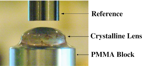
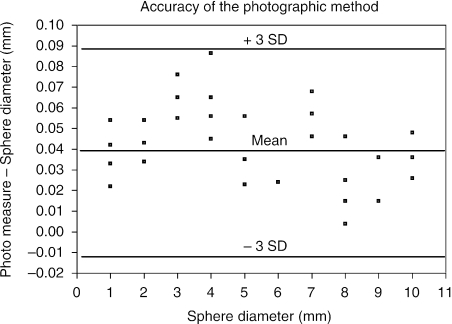
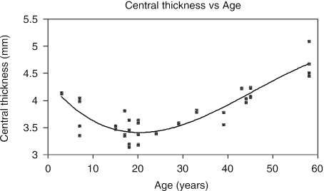
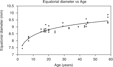
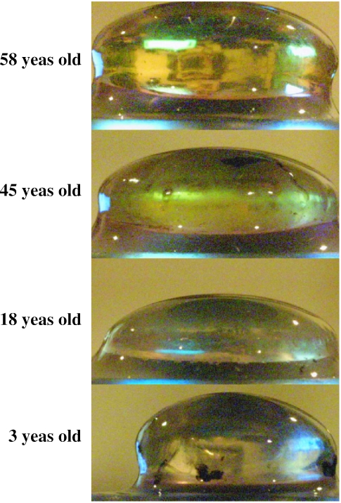
The purpose of the study was to demonstrate and evaluate the use of digital-image photography to measure the central thickness and equatorial diameter of whole fresh human crystalline lenses free of all zonular attachments. Forty-one human donor postmortem intact clear crystalline lenses, with a mean age of 28.5 +/- 16.4 years, were obtained by the contributing eye banks. The lenses were removed by the eye bank and shipped in Optisol-GS, a physiologic preservative storage medium, at 7 degrees C. The lenses were stored at the same temperature and in the same preservative throughout this study. This medium has been demonstrated to maintain the morphometric characteristics of epithelial cells. After freeing the lenses of all residual zonular attachments, digital photographs were obtained within an average of 20.9 +/- 13.4 h after death. The digital images were used to determine the central thickness and equatorial diameter of the crystalline lenses. By carefully calibrating the digital images and maintaining the lenses in physiological storage medium, reliable dimensional measurements were obtained. The dimensions for central thickness for each lens were compared to published, age-matched lenses, measured in vivo, and were found to duplicate these in vivo measurements reliably.
Figures
References
-
- Al-Ghoul KJ, Nordgren RK, Kuszak AJ, Freel CD, Costello MJ, Kuszak JR. Structural evidence of human nuclear fiber compaction as a function of ageing and cataractogenesis. Exp Eye. Res. 2001;72(3):199–214. Medline . - PubMed
-
- Bland JM, Altman DG. Statistical methods for assessing agreement between two methods of clinical measurement. Lancet. 1986:1. Medline . - PubMed
-
- Bluestein EC, Wilson ME, Wang XH, Rust PF, Apple DJ. Dimensions of the pediatric crystalline lens: implications for intraocular lenses in children. J. Pediatr. Ophthalmol. Strabismus. 1996;33:18–20. Medline . - PubMed
-
- Brown NP, Bron AJ. Lens Disorders: a Clinical Manual of Cataract Diagnosis. Oxford, UK: Butterworth-Heineman; 1996.
MeSH terms
LinkOut - more resources
Full Text Sources
