

doi: 10.1002/ana.410310505.
Corticotropin-releasing hormone-induced seizures in infant rats originate in the amygdala
Affiliations
- PMID: 1596084
- PMCID: PMC3153947
- DOI: 10.1002/ana.410310505
Item in Clipboard
Corticotropin-releasing hormone-induced seizures in infant rats originate in the amygdala
Ann Neurol.
1992 May.
Abstract
The neuroanatomical substrate of seizures induced by picomolar amounts of corticotropin-releasing hormone in infant rats was investigated. Electrographic and behavioral phenomena were monitored in 42 rat pups aged 5 to 22 days. Rat pups carried bipolar electrodes implanted in subcortical limbic structures, as well as cortical electrodes and intracerebroventricular cannulae. The administration of corticotropin-releasing hormone produced age-specific seizures within minutes, which correlated with rhythmic amygdala discharges. Paroxysmal hippocampal and cortical discharges developed subsequently in some rats. Corticotropin-releasing hormone-induced electrographic and behavioral seizures originate in the amygdala.
Figures
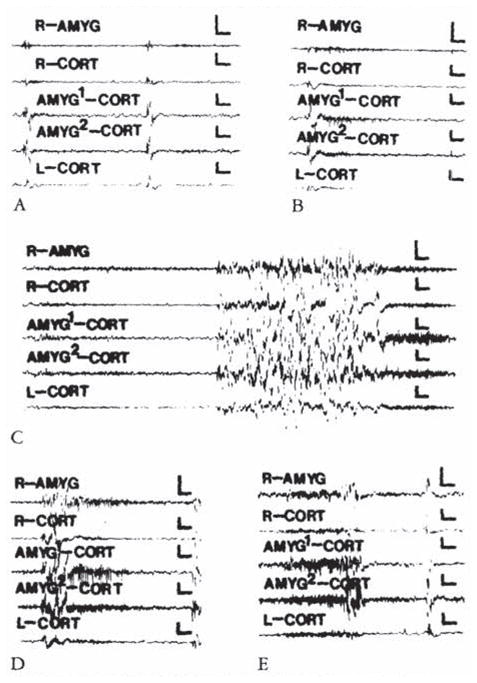
Electroencephalograms of a 5-day-old rat. (A) Before corticotropin-releasing hormone (CRH) infusion. (B, C, D, and E) Two, 11.5, 20, 59. and 85 minutes after infusion of 0.15 nmol of CRH into the cerebral ventricle. AMYC = amygdala; CORT = cortex; AMYG1/AMYG2 =one of the wires of the bipolar amygdala electrode. R/L = right / left. Vertical bar = 50 μV; horizontal bar = 1 second.
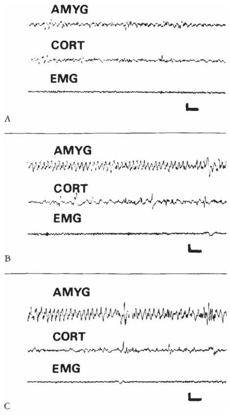
Electroencephalograms of a 9-day-old rat before corticotropin-releasing hormone (CRH) administration (A), and 9 and 120 minutes after intracerebroventricular infusion of 015 nm of CRH (B, C). The onset of semirhythmic slow wave discharges, confined to amygdala leads (AMYG). is evident in B. They persisted intermittently for several hours (C). CORT = cortical lead; EMG = motion-detecting electrodes placed over the angle of the jaw. Vertical bar = 50 μV; horizontal bar = 1 second.
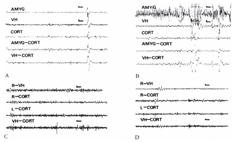
(A, B) Electroencephalograms (EEGs) of an 8-day-old infant rat. carrying both bipolar amygdala (AMYG) and ventral hippocampus (VH) electrodes, before administration of corticotropin-releasing hormone (CRH) (A) and 8 minutes after administration of 0.15 nm of the peptide (B). Paroxysmal discharges originate in the amygdala leads. Movement artifacts are later seen in all leads. (C, D) EEGs of an 11-day-old rat before (C). and 26 minutes subsequent to the administration of 0.15 nm of CRH. Though the pup displayed jaw myoclonus, only attenuation of the theta rhythm is seen in the ventral hippocampus (VH). CORT = cortex; AMYG-CORT/VH-CORT = a lead combining one of the subcortical electrodes to the cortical one. Vertical bar = 50 μV; horizontal bar = 1 second.
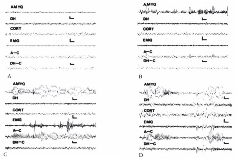
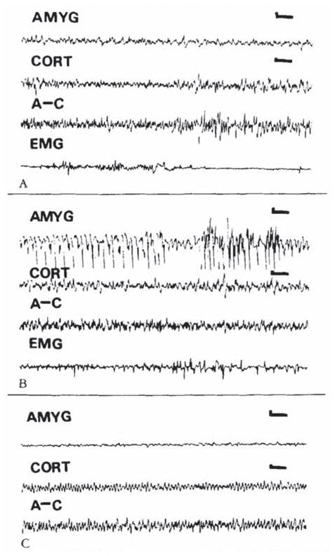
Electroencephalograms of a 13-day-old rat. (A) Baseline. (B. C. and D) Two. 35. and 174 minutes after intracerebro-ventricular administration of 0.15 nm of corticotropin-releasing hormone. High-voltage, repetitive discharges are present exclusively in bipolar amygdala (AMYG) and amygdala-cortex (A–C) leads, and not in bipolar hippocampal (DH). DH–cortex (DH–C), or the motion-detecting (EMG) leads. Vertical bar = 50 μV; horizontal bar = 1 second.
Electroencephalograms (EEGs) of an 18-day-old rat. (A) Baseline. (B. C) Nine and 143 minutes after intracerebroventrkular administration of cortkotropin-releasing hormone (0.3 nm). High-voltage spike and slow wave discharges confined to the amygdala (latency. 4 minutes) (B). Two hours later, amygdala EEG has normalized (C). AMYG = amygdala; CORT = cortex; A–C = amygdala–cortex lead; EMG = motion-detecting electrode (see text). Vertical bar = 50 μV; horizontal bar = 1 second.
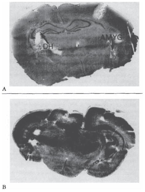
Histological verification of electrode placement in subcortical structures of the infant rat, Cresyl blue–stained. 20-μm-thick coronal sections. (A) Amygdala (AMYG) and dorsal hippocampus (DH) (B) Ventral hippocampus (VH).
References
-
- Vale W, Spiess J, Rivier C, Rivier J. Characterization of a 41-residue ovine hypothalamic peptide that stimulates secretion of corticotropin and beta-endorphin. Science. 1981;213:1394–1397. - PubMed
-
- Vale W, Rivier C, Brown MR, et al. Chemical and biological characterization of corticotropin releasing factor. Recent Prog Horm Res. 1983;83:245–270. - PubMed
-
- Koob GF, Britton KT. Behavioral effects of corticotropin-releasing factor. In: De Souza EB, Nemeroff CB, editors. Corticotropin-releasing factor: basic and clinical studies of a neuropeptide. Boca Raton: CRC; 1990. pp. 253–265.
-
- Valentino RJ, Foote SL, Aston-Jones G. Corticotropin releasing factor activates neurons of the locus coeruleus. Brain Res. 1983;270:363–367. - PubMed
-
- Siggins GR. Electrophysiology of corticotropin-releasing factor in nervous tissue. In: De Souza EB, Nemeroff CB, editors. Corticotropin-releasing factor: basic and clinical studies of a neuropeptide. Boca Raton: CRC; 1990. pp. 205–214.
