

Age effects on atrophy rates of entorhinal cortex and hippocampus
- PMID: 15961190
- PMCID: PMC1779763
- DOI: 10.1016/j.neurobiolaging.2005.03.021
Age effects on atrophy rates of entorhinal cortex and hippocampus
Abstract
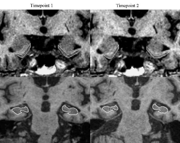
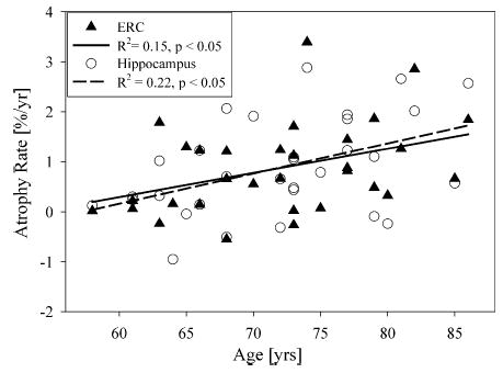
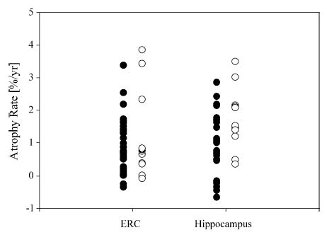
The effects of age, subcortical vascular disease, apolipoprotein E (APOE) epsilon4 allele and hypertension on entorhinal cortex (ERC) and hippocampal atrophy rates were explored in a longitudinal MRI study with 42 cognitively normal (CN) elderly subjects from 58 to 87 years old. The volumes of the ERC, hippocampus, and white matter hyperintensities (WMH) and the presence of lacunes were assessed on MR images. Age was significantly associated with increased atrophy rates of 0.04+/-0.02% per year for ERC and 0.05+/-0.02% per year for hippocampus. Atrophy rates of hippocampus, but not that of ERC increased with presence of lacunes, in addition to age. WMH, APOE epsilon4 and hypertension had no significant effect on atrophy rates. In conclusion, age and presence of lacunes should be taken into consideration in imaging studies of CN subjects and AD patients to predict AD progression and assess the response to treatment trials.
Figures
Similar articles
-
Brain aging and its modifiers: insights from in vivo neuromorphometry and susceptibility weighted imaging.Ann N Y Acad Sci. 2007 Feb;1097:84-93. doi: 10.1196/annals.1379.018. Ann N Y Acad Sci. 2007. PMID: 17413014 Free PMC article. Review.
-
White matter lesions are associated with cortical atrophy more than entorhinal and hippocampal atrophy.Neurobiol Aging. 2005 Apr;26(4):553-9. doi: 10.1016/j.neurobiolaging.2004.05.002. Neurobiol Aging. 2005. PMID: 15653183
-
Higher atrophy rate of entorhinal cortex than hippocampus in AD.Neurology. 2004 Feb 10;62(3):422-7. doi: 10.1212/01.wnl.0000106462.72282.90. Neurology. 2004. PMID: 14872024 Free PMC article.
-
Loss of entorhinal cortex and hippocampal volumes compared to whole brain volume in normal aging: the SMART-Medea study.Psychiatry Res. 2012 Jul 30;203(1):31-7. doi: 10.1016/j.pscychresns.2011.12.002. Epub 2012 Aug 19. Psychiatry Res. 2012. PMID: 22910574
-
Effects of subcortical ischemic vascular dementia and AD on entorhinal cortex and hippocampus.Neurology. 2002 Jun 11;58(11):1635-41. doi: 10.1212/wnl.58.11.1635. Neurology. 2002. PMID: 12058091 Free PMC article.
Cited by
-
Longitudinal Characterization and Biomarkers of Age and Sex Differences in the Decline of Spatial Memory.Front Aging Neurosci. 2020 Feb 20;12:34. doi: 10.3389/fnagi.2020.00034. eCollection 2020. Front Aging Neurosci. 2020. PMID: 32153384 Free PMC article.
-
Magnetic Resonance Imaging Measurement of Entorhinal Cortex in the Diagnosis and Differential Diagnosis of Mild Cognitive Impairment and Alzheimer's Disease.Brain Sci. 2021 Aug 26;11(9):1129. doi: 10.3390/brainsci11091129. Brain Sci. 2021. PMID: 34573151 Free PMC article.
-
Brain aging and its modifiers: insights from in vivo neuromorphometry and susceptibility weighted imaging.Ann N Y Acad Sci. 2007 Feb;1097:84-93. doi: 10.1196/annals.1379.018. Ann N Y Acad Sci. 2007. PMID: 17413014 Free PMC article. Review.
-
Consistent neuroanatomical age-related volume differences across multiple samples.Neurobiol Aging. 2011 May;32(5):916-32. doi: 10.1016/j.neurobiolaging.2009.05.013. Epub 2009 Jun 30. Neurobiol Aging. 2011. PMID: 19570593 Free PMC article.
-
A longitudinal study of age- and gender-related annual rate of volume changes in regional gray matter in healthy adults.Hum Brain Mapp. 2013 Sep;34(9):2292-301. doi: 10.1002/hbm.22067. Epub 2012 Mar 22. Hum Brain Mapp. 2013. PMID: 22438299 Free PMC article.
References
-
- Blacker D, Tanzi RE. The genetics of Alzheimer disease: current status and future prospects. Arch Neurol. 1998;55:294–6. - PubMed
-
- Brayne C, Gill C, Paykel ES, Huppert F, O’Connor DW. Cognitive decline in an elderly population—a two wave study of change. Psychol Med. 1995;25:673–83. - PubMed
-
- Cardenas VA, Ezekiel F, Di SV, Gomberg B, Fein G. Reliability of tissue volumes and their spatial distribution for segmented magnetic resonance images. Psychiatry Res. 2001;106:193–205. - PubMed
-
- Cervos-Navarro J, Diemer NH. Selective vulnerability in brain hypoxia. Crit Rev Neurobiol. 1991;6:149–82. - PubMed
-
- Coffey CE, Wilkinson WE, Parashos IA, Soady SA, Sullivan RJ, Patterson LJ, et al. Quantitative cerebral anatomy of the aging human brain: a cross-sectional study using magnetic resonance imaging. Neurology. 1992;42:527–36. - PubMed
Publication types
MeSH terms
Substances
Grants and funding
LinkOut - more resources
Full Text Sources
Medical
Miscellaneous
