. 2005 Aug 15;567(Pt 1):67-78.
doi: 10.1113/jphysiol.2005.089342. Epub 2005 Jun 16.

The involvement of Cav3.2/alpha1H T-type calcium channels in excitability of mouse embryonic primary vestibular neurones

Affiliations

The involvement of Cav3.2/alpha1H T-type calcium channels in excitability of mouse embryonic primary vestibular neurones

Laurence Autret et al. J Physiol. .

Abstract

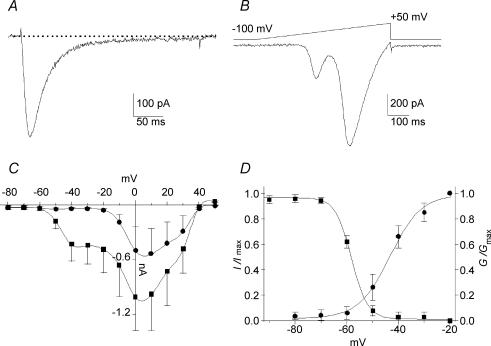
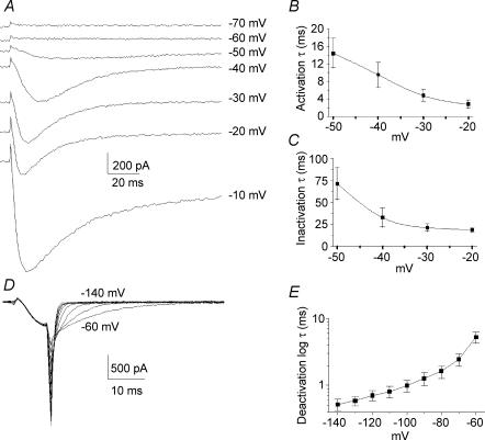
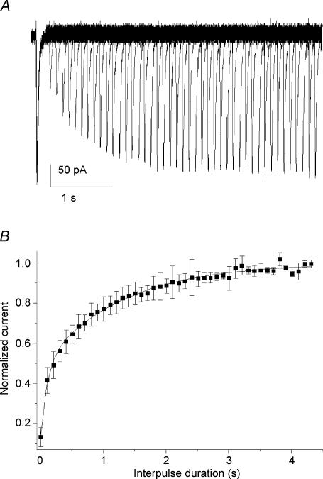
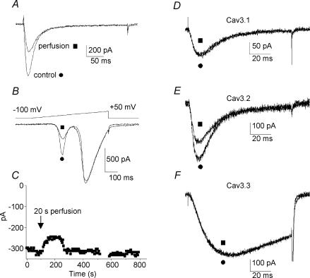
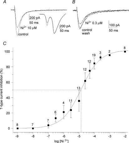
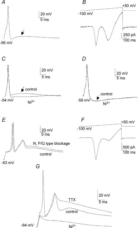
Ca2+ influx through voltage-gated calcium channels probably influences neuronal ontogenesis. Many developing neurones transiently express T-type/Cav3 calcium channels that contribute to their electrical activity and potentially to their morphological differentiation. Here we have characterized the electrophysiological properties and the functional role of a large T-type calcium current that is present in mouse developing primary vestibular neurones at embryonic day E17. This T-type current showed fast activation and inactivation, as well as slow deactivation kinetics. The overlap of activation and inactivation parameters produced a window current between -65 and -45 mV. Recovery from short-term inactivation was slow suggesting the presence of the Cav3.2 subunit. This T-type current was blocked by micromolar concentrations of Ni2+ and was inhibited by fast perfusion velocities in a similar fashion to recombinant Cav3.2 T-type channels expressed in HEK-293 cells. More importantly, current clamp experiments have revealed that the T-current could elicit afterdepolarization potentials during the repolarization phase of action potentials, and occasionally generate calcium spikes. Taken together, we demonstrate that the Cav3.2 subunit is likely to be the main T-type calcium channel subunit expressed in embryonic vestibular neurones and should play a key role in the excitability of these neurones during the ontogenesis of vestibular afferentation.


Figures

Figure 1
Figure 1. Calcium currents recorded in E17 primary vestibular neurones
A, typical trace of T-type current recorded in a neurone displaying large current amplitude. The current was evoked by a −35 mV pulse, at voltage eliciting the maximum amplitude determined during a voltage ramp. B, corresponding voltage ramp from −100 to +50 mV (640 ms duration). C, IV relationships of global calcium current recorded from Vh–100 mV from −80 to +50 mV (▪, peak current; •, sustained current). D, steady-state activation (•) and inactivation (▪) curves of T-current. The continuous lines represent the best-fits of the Boltzmann distribution. The overlap of activation and inactivation suggests a window current between −65 and −45 mV.
Figure 2
Figure 2. Activation, inactivation and deactivation properties of T-type current
A, original traces showing calcium currents at various potentials between −70 and −10 mV. The T-type current activated between −60 and −50 mV while high-voltage-activated (HVA) currents activated below −20 mV. B, time constants of activation of T-type current as function of the voltage. C, time constants of inactivation of T-type current as a function of the voltage. Both time constants of activation and inactivation were voltage dependent and present an acceleration of between −50 and −20 mV. D, original traces showing tail currents obtained by deactivating pulses from −140 mV to −60 mV. Pipette and cell capacitances were corrected and series resistances reduced to about 85%. E, plot of mean deactivating time constants of the fit of the tail currents as a function of the voltage.
Figure 3
Figure 3. Time dependence of recovery following short inactivation
A, superimposed current traces obtained in an E17 vestibular neurone showing the current growth when the interval of two paired pulses (100 ms, −42 mV) increased from 0 ms up to 4.4 s (step 100 ms). B, plot of the mean normalized current amplitudes as a function of the interpulse interval. The relationships were fitted with two exponentials (continuous line). The time constants were 102 ± 13 and 1281 ± 41 ms, and amplitudes 0.38 ± 0.03 and 0.53 ± 0.02 (n = 22) for the fast and slow components, respectively.
Figure 4
Figure 4. Mechanical responses of T-type current to the extracellular recording medium perfusion
A, original trace of T-type current recorded in E17 vestibular neurone, before (•) and after (▪) application of extracellular recording medium at a velocity of 1 μl s−1. B, corresponding voltage ramps showing the absence of shift in I–V relationship. Note that HVA calcium currents were not reduced. C, long duration recordings showing the reversibility of the extracellular medium perfusion effect on vestibular neurones. Application of recording medium (20 μl, 20 s) induced a current decrease, which reached a minimum in about 60 s and was followed by an amplitude increase to return to the initial amplitude after about 220 s. D–F, T-currents recorded in HEK-293 cells transfected with the different Cav3/α1 T-channel subunits. Trace currents were illustrated before (•) and after (▪) application of extracellular recording medium at a velocity of 1 μl s−1. While Cav3.1/α1G (D) and Cav3.3/α1I (F) were insensitive to the perfusion, Cav3.2/α1H (E) decreased by about 30%.
Figure 5
Figure 5. Effect of increasing concentration of Ni2+
A, effect of 10 μm Ni2+ application on T-current. The HVA calcium currents were not affected (inset). B, reversible effect of 0.3 μm Ni2+ application on T-current. C, concentration dependence of the blockade of T-type current by Ni2+. The smooth curve represents the fit of the Hill equation revealing an IC50 of 13.4 μm. The Hill coefficient was 0.7 ± 0.1. Dotted lines illustrate the IC50. The number of observations is given above the error bars. The mechanosensitivity response is not reported in the curve.
Figure 6
Figure 6. Implication of T-type current in AP profiles
A, action potential recorded in current clamp showing an afterdepolarization potential (ADP) (arrow) following the repolarization phase. B, corresponding voltage ramp recorded in the same neurone after application of TTX (300 nm), TEA (60 mm) and 4-AP (5 mm) to block the Na+ currents and limit K+ conductances. The voltage ramp reveals the presence of a large T-type current. It is noteworthy that the remaining K+ conductances were presented at high voltage, mainly due to the absence of CsCl in the internal pipette medium. C, action potential presenting an ADP (arrow) which was blocked by the application of 10 μm Ni2+. D, action potential presenting an afterhyperpolarization (AHP) that was insensitive to 30 μm Ni2+ application. E, Na+-dependent action potentials followed by Ca2+ spikes. Application of ω-conotoxin GVIA (500 nm) and ω-agatoxin IVA (300 nm) to block N and P/Q HVA calcium channels, respectively, did not change the calcium spike amplitude. F, voltage ramp recorded in the same neurone showing a high amplitude T-type current after blockage of the Na+ and K+ conductances. In the presence of GVIA and ω-agatoxin IVA, the remaining HVA component revealed the L- and R-type calcium currents that contribute to a small fraction of the global calcium current in primary vestibular neurones. G, Na+-dependent action potential followed by a Ca2+ spike. Application of TTX (300 nm) eliminated the Na+-dependent action potential without affecting the calcium spike which was completely abolished by the addition of 30 μm Ni2+.

References

    1. Anniko M. Early development and maturation of the spiral ganglion. Acta Otolaryngol. 1983;95:263–276. - PubMed
    1. Baird RA, Desmadryl G, Fernandez C, Goldberg JM. The vestibular nerve of the chinchilla. II. Relation between afferent response properties and peripheral innervation patterns in the semicircular canals. J Neurophysiol. 1988;60:182–203. - PubMed
    1. Ben-Tabou S, Keller E, Nussinovitch I. Mechanosensitivity of voltage-gated calcium currents in rat anterior pituitary cells. J Physiol. 1994;476:29–39. - PMC - PubMed
    1. Berthier C, Monteil A, Lory P, Strube C. α1H mRNA in single skeletal muscle fibres accounts for T-type calcium current transient expression during fetal development in mice. J Physiol. 2002;539:681–691. - PMC - PubMed
    1. Bijlenga P, Liu JH, Espinos E, Haenggeli CA, Fischer-Lougheed J, Bader CR, Bernheim L. T-type alpha 1H Ca2+ channels are involved in Ca2+ signaling during terminal differentiation (fusion) of human myoblasts. Proc Natl Acad Sci U S A. 2000;97:7627–7632. - PMC - PubMed

Publication types

LinkOut - more resources