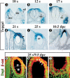
Wnt-dependent regulation of inner ear morphogenesis is balanced by the opposing and supporting roles of Shh
- PMID: 15961523
- PMCID: PMC1172066
- DOI: 10.1101/gad.1303905
Wnt-dependent regulation of inner ear morphogenesis is balanced by the opposing and supporting roles of Shh
Abstract
The inner ear is partitioned along its dorsal/ventral axis into vestibular and auditory organs, respectively. Gene expression studies suggest that this subdivision occurs within the otic vesicle, the tissue from which all inner ear structures are derived. While the specification of ventral otic fates is dependent on Shh secreted from the notochord, the nature of the signal responsible for dorsal otic development has not been described. In this study, we demonstrate that Wnt signaling is active in dorsal regions of the otic vesicle, where it functions to regulate the expression of genes (Dlx5/6 and Gbx2) necessary for vestibular morphogenesis. We further show that the source of Wnt impacting on dorsal otic development emanates from the dorsal hindbrain, and identify Wnt1 and Wnt3a as the specific ligands required for this function. The restriction of Wnt target genes to the dorsal otocyst is also influenced by Shh. Thus, a balance between Wnt and Shh signaling activities is key in distinguishing between vestibular and auditory cell types.
Figures


Similar articles
-
Specification of the mammalian cochlea is dependent on Sonic hedgehog.Genes Dev. 2002 Sep 15;16(18):2365-78. doi: 10.1101/gad.1013302. Genes Dev. 2002. PMID: 12231626 Free PMC article.
-
Opposing gradients of Gli repressor and activators mediate Shh signaling along the dorsoventral axis of the inner ear.Development. 2007 May;134(9):1713-22. doi: 10.1242/dev.000760. Epub 2007 Mar 29. Development. 2007. PMID: 17395647
-
The cochlear sensory epithelium derives from Wnt responsive cells in the dorsomedial otic cup.Dev Biol. 2015 Mar 1;399(1):177-187. doi: 10.1016/j.ydbio.2015.01.001. Epub 2015 Jan 12. Dev Biol. 2015. PMID: 25592224 Free PMC article.
-
Signaling cascade coordinating growth of dorsal and ventral tissues of the vertebrate brain, with special reference to the involvement of Sonic Hedgehog signaling.Anat Sci Int. 2005 Mar;80(1):30-6. doi: 10.1111/j.1447-073x.2005.00096.x. Anat Sci Int. 2005. PMID: 15794128 Review.
-
Hearing crosstalk: the molecular conversation orchestrating inner ear dorsoventral patterning.Wiley Interdiscip Rev Dev Biol. 2018 Jan;7(1):10.1002/wdev.302. doi: 10.1002/wdev.302. Epub 2017 Oct 11. Wiley Interdiscip Rev Dev Biol. 2018. PMID: 29024472 Free PMC article. Review.
Cited by
-
Development and evolution of the vestibular apparatuses of the inner ear.J Anat. 2021 Oct;239(4):801-828. doi: 10.1111/joa.13459. Epub 2021 May 28. J Anat. 2021. PMID: 34047378 Free PMC article. Review.
-
Elongation factor 1 alpha1 and genes associated with Usher syndromes are downstream targets of GBX2.PLoS One. 2012;7(11):e47366. doi: 10.1371/journal.pone.0047366. Epub 2012 Nov 8. PLoS One. 2012. PMID: 23144817 Free PMC article.
-
Divergent roles for Wnt/β-catenin signaling in epithelial maintenance and breakdown during semicircular canal formation.Development. 2013 Apr;140(8):1730-9. doi: 10.1242/dev.092882. Epub 2013 Mar 13. Development. 2013. PMID: 23487315 Free PMC article.
-
Key Genes and Pathways Associated With Inner Ear Malformation in SOX10 p.R109W Mutation Pigs.Front Mol Neurosci. 2018 Jun 5;11:181. doi: 10.3389/fnmol.2018.00181. eCollection 2018. Front Mol Neurosci. 2018. PMID: 29922125 Free PMC article.
-
Fgf3 and Fgf16 expression patterns define spatial and temporal domains in the developing chick inner ear.Brain Struct Funct. 2017 Jan;222(1):131-149. doi: 10.1007/s00429-016-1205-1. Epub 2016 Mar 19. Brain Struct Funct. 2017. PMID: 26995070 Free PMC article.
References
-
- Acampora D., Merlo, G.R., Paleari, L., Zerega, B., Postiglione, M.P., Mantero, S., Bober, E., Barbieri, O., Simeone, A., and Levi, G. 1999. Craniofacial, vestibular and bone defects in mice lacking the Distal-less-related gene Dlx5. Development 126: 3795–3809. - PubMed
-
- Alvarez Y., Alonso, M.T., Vendrell, V., Zelarayan, L.C., Chamero, P., Theil, T., Bosl, M.R., Kato, S., Maconochie, M., Riethmacher, D., et al. 2003. Requirements for FGF3 and FGF10 during inner ear formation. Development 130: 6329–6338. - PubMed
-
- Baas D., Bumsted, K.M., Martinez, J.A., Vaccarino, F.M., Wikler, K.C., and Barnstable, C.J. 2000. The subcellular localization of Otx2 is cell-type specific and developmentally regulated in the mouse retina. Brain Res. Mol. Brain Res. 78: 26–37. - PubMed
-
- Barald K.F. and Kelley, M.W. 2004. From placode to polarization: New tunes in inner ear development. Development 131: 4119–4130. - PubMed
-
- Bienz M. and Clevers, H. 2003. Armadillo/β-catenin signals in the nucleus-proof beyond a reasonable doubt? Nat. Cell Biol. 5: 179–182. - PubMed
Publication types
MeSH terms
Substances
Grants and funding
LinkOut - more resources
Full Text Sources
Other Literature Sources
Molecular Biology Databases
Research Materials
