. 2005 Jul 6;24(13):2472-80.
doi: 10.1038/sj.emboj.7600717. Epub 2005 Jun 16.

Immunodetection of disease-associated mutant PrP, which accelerates disease in GSS transgenic mice

Affiliations

Immunodetection of disease-associated mutant PrP, which accelerates disease in GSS transgenic mice

Karah E Nazor et al. EMBO J. .

Abstract

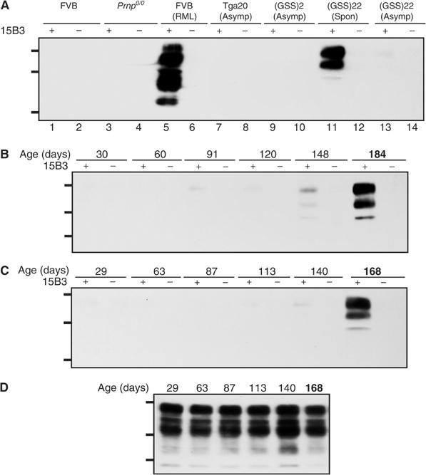
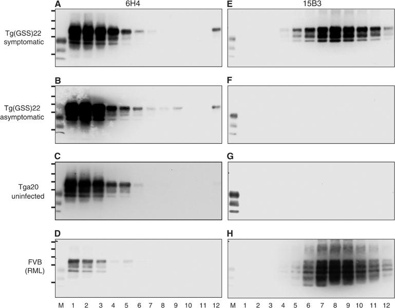
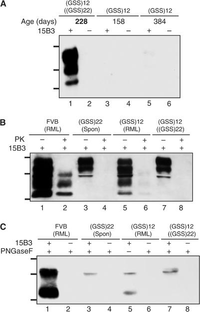
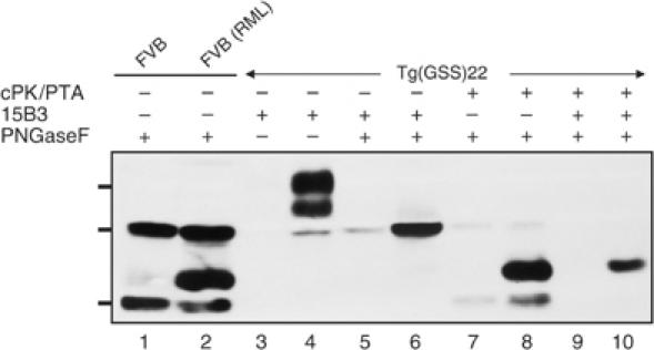
The absence of infectivity-associated, protease-resistant prion protein (PrP(Sc)) in the brains of spontaneously sick transgenic (Tg) mice overexpressing PrP linked to Gerstmann-Sträussler Scheinker syndrome, and the failure of gene-targeted mice expressing such PrP to develop disease spontaneously, challenged the concept that mutant PrP expression led to spontaneous prion production. Here, we demonstrate that disease in overexpressor Tg mice is associated with accumulation of protease-sensitive aggregates of mutant PrP that can be immunoprecipitated by the PrP(Sc)-specific monoclonal antibody designated 15B3. Whereas Tg mice expressing multiple transgenes exhibited accelerated disease when inoculated with disease-associated mutant PrP, Tg mice expressing mutant PrP at low levels failed to develop disease either spontaneously or following inoculation. These studies indicate that inoculated mutant PrP from diseased mice promotes the aggregation and accumulation of pre-existing pathological forms of mutant PrP produced as a result of transgene overexpression. Thus, while pathological mutant PrP possesses a subset of PrP(Sc) characteristics, we now show that the attribute of prion transmission suggested by previous studies is more accurately characterized as disease acceleration.


Figures

Figure 1
Figure 1
Spontaneous disease in Tg(GSS) mice correlates with the accumulation of 15B3-reactive MoPrP-P101L. (A) Brain extracts were treated with anti-mouse IgM-coupled magnetic beads in the presence or absence of Mab 15B3 as indicated and immunoprecipitates were analyzed by SDS–PAGE and Western blotting as described. Lanes 1 and 2, uninoculated FVB mouse; lanes 3 and 4, Prnp0/0 mouse; lanes 5 and 6, FVB mouse inoculated with mouse-adapted RML scrapie prions; lanes 7 and 8, asymptomatic Tga20 mouse; lanes 9 and 10, asymptomatic Tg(GSS)2 mouse killed at 572 days of age; lanes 11 and 12, spontaneously sick Tg(GSS)22 mouse killed at 188 days of age; lanes 13 and 14, asymptomatic Tg(GSS)22 mouse killed at 30 days of age. (B, C) Kinetics of disease-associated MoPrP-P101L accumulation in Tg(GSS)22 mice. Brain tissues were collected from two independent birth cohorts of Tg(GSS)22 mice killed at various ages, as indicated. The second cohort of Tg(GSS)22 mice was established approximately 1 year after the first. Brain extracts were treated with anti-mouse IgM-coupled magnetic beads in the presence or absence of Mab 15B3 as indicated and immunoprecipitates were analyzed by SDS–PAGE and Western blotting using HRP-conjugated anti-PrP antibody 6H4. (D) Equal amounts of protein in the brains of Tg(GSS)22 mice killed at different ages, as indicated, were analyzed by SDS–PAGE and Western blotting. In (B–D), the ages of asymptomatic Tg(GSS)22 mice are shown in plain text, while mice in which clinical symptoms of disease were manifest at the time of killing are indicated by bold type. The positions of protein molecular weight markers of 35.5, 28.8, and 22.0 kDa from top to bottom are shown.
Figure 2
Figure 2
Characterization of MoPrP-P101L aggregates in spontaneously sick Tg(GSS) mice. Sucrose gradient fractionation of brain homogenates followed by immunoprecipitation with Mab 6H4 (A–D) or 15B3 (E–H). (A, E) Spontaneously sick Tg(GSS)22 mouse killed at 181 days of age; (B, F) healthy Tg(GSS)22 mouse killed at 30 days of age; (C, G) uninoculated Tga20 mouse; (D, H) FVB mouse inoculated with mouse-adapted RML scrapie prions. Immunoprecipitated samples were analyzed by SDS–PAGE and Western blotting using HRP-conjugated Mab 6H4. M=PrP27−30 from BSE-infected cow. In each case, lanes 1–12 refer to 1 ml fractions isolated from the top to the bottom of sucrose density gradients. The positions of protein molecular weight markers of 72, 55, 40, 33, and 24 kDa from top to bottom are shown.
Figure 3
Figure 3
Disease-associated MoPrP-P101L in sick Tg(GSS) mice inoculated with extracts from spontaneously sick Tg(GSS) mice and RML prions. (A) Brain extracts were treated with anti-mouse IgM-coupled magnetic beads in the presence or absence of Mab 15B3 as indicated and immunoprecipitates were analyzed by SDS–PAGE and Western blotting using HRP-conjugated Mab 6H4. Lanes 1 and 2, sick Tg(GSS)12 mouse killed at 228 days of age inoculated at 35 days of age with brain extract from spontaneously sick Tg(GSS)22 mouse, brain 1; lanes 3 and 4, asymptomatic uninoculated Tg(GSS)12 mouse, killed at 158 days of age; lanes 5 and 6, asymptomatic uninoculated Tg(GSS)12 mouse, killed at 384 days of age. The ages of asymptomatic Tg(GSS)12 mice are shown in plain text, while mice in which clinical symptoms of disease were manifest at the time of killing are indicated by bold type. (B) Following immunoprecipitation with Mab 15B3, immune complexes were treated with 20 μg/ml PK for 1 h at 37°C as indicated and analyzed by SDS–PAGE and Western blotting. (C) Following immunoprecipitation with Mab 15B3, immune complexes were treated with PNGase F as indicated and analyzed by SDS–PAGE and Western blotting. Samples in (B, C) are equivalent and are as follows: lanes 1 and 2, sick FVB mouse inoculated with mouse-adapted RML scrapie prions; lanes 3 and 4, spontaneously sick Tg(GSS)22 mouse killed at 159 days of age; lanes 5 and 6, sick Tg(GSS)12 mouse killed at 211 days following inoculation with mouse-adapted RML scrapie prions; lanes 7 and 8, sick Tg(GSS)12 mouse killed at 193 days following inoculation with brain extract from a spontaneously sick Tg(GSS)22 mouse. The positions of protein molecular weight markers of 35.5, 28.8, and 22 kDa from top to bottom are shown.
Figure 4
Figure 4
Relative specificities of NaPTA precipitation combined with ‘cold PK' treatment and 15B3 immunoprecipitation for pathological MoPrP-P101L. Lane 1, brain extract from uninfected FVB mouse digested with PNGase F; lane 2, brain extract of sick RML-inoculated FVB mouse digested with PNGase F; lane 3, brain extract from an asymptomatic Tg(GSS)22 mouse immunoprecipitated with Mab 15B3; lane 4, brain extract from a sick Tg(GSS)22 mouse immunoprecipitated with Mab 15B3; lane 5: brain extract from an asymptomatic Tg(GSS)22 mouse immunoprecipitated with Mab 15B3 and digested with PNGase F; lane 6, brain extract from a sick Tg(GSS)22 mouse immunoprecipitated with Mab 15B3 and digested with PNGase F; lane 7, ‘cold PK' digestion followed by NaPTA precipitation, referred to as cPK/PTA, and PNGase digestion of brain extract from an asymptomatic Tg(GSS)22 mouse; lane 8, ‘cold PK' digestion, NaPTA precipitation, and PNGase digestion of brain extract from a sick Tg(GSS)22 mouse; lane 9, ‘cold PK' digestion, NaPTA precipitation, Mab 15B3 immunoprecipitation, and PNGase digestion of brain extract from an asymptomatic Tg(GSS)22 mouse; lane 10, ‘cold PK' digestion, NaPTA precipitation, Mab 15B3 immunoprecipitation, and PNGase digestion of brain extract from a sick Tg(GSS)22 mouse. Samples were analyzed by SDS–PAGE and Western blotting using HRP-conjugated Mab 6H4. The asymptomatic Tg(GSS)22 mouse was killed at 30 days of age while the sick Tg(GSS)22 was killed at 188 days of age. The positions of protein molecular weight markers of 35.5, 28.8, and 22 kDa from top to bottom are shown.

References

    1. Bessen RA, Marsh RF (1992) Biochemical and physical properties of the prion protein from two strains of the transmissible mink encephalopathy agent. J Virol 66: 2096–2101 - PMC - PubMed
    1. Bessen RA, Marsh RF (1994) Distinct PrP properties suggest the molecular basis of strain variation in transmissible mink encephalopathy. J Virol 68: 7859–7868 - PMC - PubMed
    1. Brown P, Gibbs CJ Jr, Rodgers-Johnson P, Asher DM, Sulima MP, Bacote A, Goldfarb LG, Gajdusek DC (1994) Human spongiform encephalopathy: the National Institutes of Health series of 300 cases of experimentally transmitted disease. Ann Neurol 35: 513–529 - PubMed
    1. Chandler RL (1961) Encephalopathy in mice produced by inoculation with scrapie brain material. Lancet 1: 1378–1379 - PubMed
    1. Chen SG, Teplow DB, Parchi P, Teller JK, Gambetti P, Autilio-Gambetti L (1995) Truncated forms of the human prion protein in normal brain and in prion diseases. J Biol Chem 270: 19173–19180 - PubMed

Publication types