. 2006 Jan;13(1):41-52.
doi: 10.1038/sj.cdd.4401699.

Intravenous apoptotic spleen cell infusion induces a TGF-beta-dependent regulatory T-cell expansion

Affiliations

Intravenous apoptotic spleen cell infusion induces a TGF-beta-dependent regulatory T-cell expansion

F Kleinclauss et al. Cell Death Differ. 2006 Jan.

Abstract

Apoptotic leukocytes are endowed with immunomodulatory properties that can be used to enhance hematopoietic engraftment and prevent graft-versus-host disease (GvHD). This apoptotic cell-induced tolerogenic effect is mediated by host macrophages and not recipient dendritic cells or donor phagocytes present in the bone marrow graft as evidenced by selective cell depletion and trafficking experiments. Furthermore, apoptotic cell infusion is associated with TGF-beta-dependent donor CD4+CD25+ T-cell expansion. Such cells have a regulatory phenotype (CD62L(high) and intracellular CTLA-4+), express high levels of forkhead-box transcription factor p3 (Foxp3) mRNA and exert ex vivo suppressive activity through a cell-to-cell contact mechanism. In vivo CD25 depletion after apoptotic cell infusion prevents the apoptotic cell-induced beneficial effects on engraftment and GvHD occurrence. This highlights the role of regulatory T cells in the tolerogenic effect of apoptotic cell infusion. This novel association between apoptosis and regulatory T-cell expansion may also contribute to preventing deleterious autoimmune responses during normal turnover.


Figures
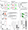
Figure 1
Figure 1
Recipient macrophages are mainly involved in the graft-facilitating effect of apoptotic spleen cell infusion. (a) Green-PKH- or CFSE-labeled apoptotic cells (detectable in the FL1 channel) were used to explore phagocytes involved in the apoptotic cell uptake. Left panel: 45 min after BMT, fluorescent apoptotic cells infused simultaneously with BM graft were found in splenic DC (F4/80lowCD11c+FL1+; white arrows) as well as in macrophages (F4/80highCD11cFL1+; black arrows). Results from a representative experiment out of 4. Middle panel: non-fluorescent apoptotic cells used as controls. Right panel: kinetics of the appearance of CFSE+CD11c+ DC (■) and CFSE+F4/80+ macrophages (●) after infusion of CFSE-labeled apoptotic cells. The percentage of CFSE+ cells contained within the indicated splenic cell population is listed on the y axis. n = 3 mice per time point. Bars represent S.D. (b) Sca-1 cell sorting was used to deplete phagocytes from the BM (left panel: a representative sorting experiment out of 4). Right panel: donor engraftment after infusion of total C57BL/6 BM (106 cells) or sca-1-enriched graft (Sca-1, 105 cells) with (Apo cells, hatched bars) or without (white bars) apoptotic cells. Pooled results from 3 independent experiments (at least 20 mice/group) expressed in % of engrafted mice. * P < 0.05. (c) Clodronate-loaded liposomes were infused before BMT to deplete host splenic phagocyting cells. Left panel: flow cytometric CD11b staining shows the effective depletion of CD11b+ macrophages at day 3 after clodronate-liposome infusion. CD11b staining in PBS loaded-liposome treated mice was shown as control. Right panel: donor engraftment after infusion of total BM (106 cells) with (hatched bars) or without (white bars) apoptotic cells in mice treated by PBS-liposomes or clodronate-liposomes. (d) Recipient CD11c+ DC were depleted at day-1 before BMT using a single infusion of diphtheria toxin (DT). Top panel: a representative depletion of eGFP+CD11c+ cells after a 20 h DT treatment. Bottom panel: donor engraftment after infusion of total BM (106 cells) with (hatched bars) or without (open bars) apoptotic cells in DT-treated transgenic mice. Pooled results from 2 independent experiments expressed in % of engrafted mice. * P < 0.05.
Figure 2
Figure 2
Apoptotic spleen cell infusion simultaneously with a bone marrow graft induces an in vivo TGF-β-dependent splenic expansion of CD4+CD25+ T cells. (a) Splenic CD4+CD25+ T cells increase after apoptotic spleen cell infusion. These cells were evaluated by cytometry in different groups: naive mice, engrafted and non-engrafted mice given apoptotic cells, engrafted mice given only a higher number of BM cells (3 × 10⁶) and engrafted mice given a higher conditioning regimen (7 Gy). Boxes represent mean + SEM, bars the S.D. and circles the highest and lowest values. *: P < 0.01. (b) TGF-β neutralization inhibits apoptotic spleen cell-induced splenic CD4+CD25+ T cell increase. The percentage of splenic CD4+CD25+ T cells was evaluated 7–9 weeks post-graft in TGF-β mAb-treated mice (n = 7) vs in irrelevant isotype Ab-treated mice (n= 9). Pooled results of 2 independent experiments. *: P < 0.01.
Figure 3
Figure 3
Apoptotic spleen cell infusion simultaneously with a bone marrow graft induces in vivo splenic expansion of CD4+CD25+ regulatory T cells expressing high levels of Foxp3 mRNA and exhibiting in vitro cell-to-cell contact-dependent suppression. (a) Expanded splenic CD4+CD25+ T cells from engrafted and non-engrafted mice were characterized 7–9 weeks post-BMT. CD25, H-2d and CD62L expressions were evaluated on the CD4+CD3+ T cell gate (CD3+ ; CD4+ ; SSC vs FSC), whereas intracellular CTLA-4 expression was evaluated on the CD3+CD4+CD25+ T cell gate. (b) Seven to nine weeks post-BMT, cell-sorted splenic CD4+CD25+ T cells were analyzed for Foxp3 mRNA expression in engrafted (n = 2, hatched bars) and non-engrafted (n = 1, open bar) mice that had been given apoptotic cells plus BM cells. Relative Foxp3 mRNA expression was obtained by dividing relative Foxp3 expression in the CD4+CD25+ T cell fraction by that of the CD4+CD25 T cell fraction. (c) Foxp3 mRNA expression is assessed in whole spleen of either mice given BM alone (left open bars, n= 4), mice given apoptotic cells plus BM (hatched bars, n= 6) 7–9 week post-BMT. Foxp3 mRNA expression in the spleen of naive BALB/c mice (right open bars, n= 8) was shown as control. Results are expressed as mean ± SEM of normalized Foxp3 expression (obtained by dividing the relative amount of Foxp3 mRNA for each sample by the relative amount of GAPDH mRNA of the same sample). (d) Splenic CD4+ T cells from engrafted (left panel, n = 3) or non-engrafted (right panel, n = 2) BALB/c mice given donor FVB BM and apoptotic cells were cultured with or without allogeneic (FVB donor or C57BL/6 third party) stimulator cells. T cells were CD25-depleted (open bars) or not (hatched bars). MLR proliferation was assessed by [3H]thymidine uptake (cpm). *: P < 0.01. (e) Cell-sorted CD4+CD25+ T cells were added to a MLR between stimulator irradiated splenic FVB donor cells and CD25-depleted responder BALB/c recipient CD4+ T cells (n = 2) with or without (Ø) anti-IL10R, anti-TGF-β mAb or both. A transwell was also used to separate the MLR from sorted CD4+CD25+ T cells. Results are expressed as relative proliferation obtained by dividing the CD4+ T cell proliferation in response to allogeneic stimulators by the proliferation obtained in similar conditions but in the presence of blocking Abs or transwell. [3H]thymidine uptake > 9000 cpm. *: P < 0.05.
Figure 4
Figure 4
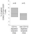
Depletion of CD11c+ dendritic cells has no effect on CD62LhighCD4+CD25+ T cell increase induced by apoptotic spleen cell infusion. Recipient CD11c+ DC were depleted at day -1 before BMT using a single infusion of diphtheria toxin (DT). CD11c/eGFP/DTR transgenic mice given donor apoptotic cells plus BM were treated (left box, n= 8) or not (right box, n= 5) with DT. CD62L+CD4+CD25+ T cells were analyzed by cytometry 7–9 weeks post-BMT. Boxes represent mean ± SEM, bars the SD.
Figure 5
Figure 5
Apoptotic spleen cell infusion simultaneously with a bone marrow graft induces an early increase of CD62L+CD4+CD25+ T cells of both host and donor origin. (a) Host macrophages are involved in the early increase of CD62L+CD4+CD25+ T cells induced by apoptotic spleen cell infusion. Splenic CD62L+CD4+CD25+ T cells were analyzed by cytometry at different time points after the infusion of FVB BM alone (□, n=5), or FVB BM plus FVB apoptotic cells (■, n=5). Such cells were also analyzed in BALB/c mice pre-treated with clodronate-liposomes and grafted with FVB BM plus FVB apoptotic cells (▲, n=3) and in irradiated BALB/c mice (△, n = 3). *: P < 0.05. (b) Percentage of donor CD62L+CD4+CD25+ T cells in the spleen of mice given FVB BM alone (□, n=5) or BM plus apoptotic spleen cells (■, n=5) at different early time points after BMT. *: P < 0.05.
Figure 6
Figure 6
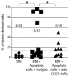
CD25+ cell depletion significantly reduces the number of circulating donor-derived cells after apoptotic spleen cell infusion. Percentage of donor-derived cells was evaluated 7–9 week post-graft in the spleen of mice given only BM (●, n=10), BM and apoptotic cells plus anti-CD25 mAb (▲, n=10) or plus irrelevant mAb (■, n=12). Number of engrafted mice in each group was shown. Dotted line indicates 15% of donor-derived cells corresponding to engraftment. Pooled results of 2 independent experiments. *: P < 0.05.
Figure 7
Figure 7
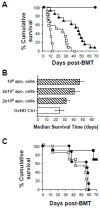
CD25+ cell depletion prevents the protecting effect of donor apoptotic spleen cell administration on graft-versus-host disease occurrence.(a) Cumulative survival was assessed in the GvHD model in mice receiving BM plus splenic T cells and donor apoptotic spleen cells (▲) or not (control GvHD group; □). Mice receiving BM alone (●) or only lethally irradiated (○) were also used as control. Results of a representative experiment out of 4 are expressed in % of cumulative survival. (b) Donor apoptotic spleen cells delays lethal GvHD occurrence in a dose-dependent manner. Median survival time was assessed in mice receiving increasing number of apoptotic cells (2 × 10⁶, 2 × 10⁷ or 108 apoptotic spleen cells; hatched bars) or not (GvHD Ctrl group; open bar) in addition to BM and splenic T cells. Pooled results from 4 independent experiments (10 mice per group, excepted for ctrl group n= 5) are expressed in median survival time (day) ± SD. (c) Delayed CD25+ cell depletion abolishes the protective effects of donor apoptotic spleen cell infusion on lethal GvHD occurrence. Cumulative survival was assessed in mice receiving BM plus splenic T cells (GvHD Ctrl group, □); with apoptotic cells and treated at day 3 and 6 by anti-CD25 mAb (●) or irrelevant IgG1 control (■). Results of a representative experiment out of 3 are expressed in % of cumulative survival. 10 mice per group.

Similar articles

Cited by

References

    1. Kamradt T, Mitchison NA. Tolerance and autoimmunity. N Engl J Med. 2001;344:655–64. - PubMed
    1. Sakaguchi S. Regulatory T cells: key controllers of immunologic self-tolerance. Cell. 2000;101:455–8. - PubMed
    1. Shevach EM. CD4+ CD25+ suppressor T cells: more questions than answers. Nat Rev Immunol. 2002;2:389–400. - PubMed
    1. Wood KJ, Sakaguchi S. Regulatory T cells in transplantation tolerance. Nat Rev Immunol. 2003;3:199–210. - PubMed
    1. Cohen JL, Trenado A, Vasey D, Klatzmann D, Salomon BL. CD4(+)CD25(+) immunoregulatory T Cells: new therapeutics for graft-versus-host disease. J Exp Med. 2002;196:401–6. - PMC - PubMed

Publication types

MeSH terms