

Detection of lymph nodes micrometastases in Dukes' A and B colorectal cancer using anti-cytokeratin antibodies AE1/AE3
- PMID: 15962393
- PMCID: PMC4315979
- DOI: 10.3748/wjg.v11.i23.3640
Detection of lymph nodes micrometastases in Dukes' A and B colorectal cancer using anti-cytokeratin antibodies AE1/AE3
Abstract
Aim: To detect lymph nodes micrometastases and analyze its correlation with clinicopathological parameters in Dukes' A and B colorectal cancer patients.
Methods: One hundred and fourteen patients with colorectal cancer (Dukes' A 16; Dukes' B 98) undergoing curative operation without histological lymph nodes metastases were studied between 2001 and 2003. A total of 2,481 lymph nodes were analyzed using monoclonal cytokeratin antibody AE1/AE3 (DAKO, Carpinteria, CA) for immunohistochemistry.
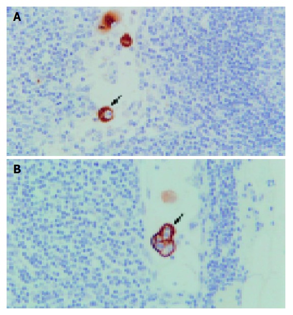
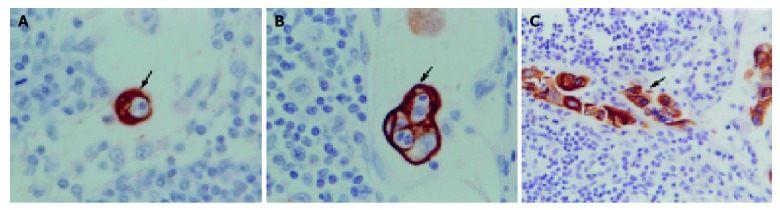
Results: In total, 33 (29%) patients were positive for cancer cell by immunohistochemistry. In 31 (94%) patients of them positive nodes showed single tumor cell or small groups of tumor cells; and tumor deposits measuring 0.2 and 0.37 mm in diameter in another 2 (6%) patients. Micrometastases were mainly located in the subcapsular sinus or paracortical sinus. There was no correlation between the positive lymph nodes and gender, age, tumor site, tumor size, histological type, histological grade, invasion depth, Dukes' staging and microsatellite instability (P>0.05).
Conclusion: Our findings suggest that immunohistochemical technique using monoclonal cytokeratin antibody AE1/AE3 may be a sensitive and reliable method for detecting lymph nodes micrometastases in Dukes' A and B colorectal cancer. The clinical significance of lymph nodes micrometastases is still not confirmed.
Figures
References
-
- Broll R, Schauer V, Schimmelpenning H, Strik M, Woltmann A, Best R, Bruch HP, Duchrow M. Prognostic relevance of occult tumor cells in lymph nodes of colorectal carcinomas: an immunohistochemical study. Dis Colon Rectum. 1997;40:1465–1471. - PubMed
-
- Hermanek P. Disseminated tumor cells versus micrometastasis: definitions and problems. Anticancer Res. 1999;19:2771–2774. - PubMed
-
- Natsugoe S, Mueller J, Stein HJ, Feith M, Höfler H, Siewert JR. Micrometastasis and tumor cell microinvolvement of lymph nodes from esophageal squamous cell carcinoma: frequency, associated tumor characteristics, and impact on prognosis. Cancer. 1998;83:858–866. - PubMed
-
- Adell G, Boeryd B, Frånlund B, Sjödahl R, Håkansson L. Occurrence and prognostic importance of micrometastases in regional lymph nodes in Dukes' B colorectal carcinoma: an immunohistochemical study. Eur J Surg. 1996;162:637–642. - PubMed
-
- Occult axillary lymph-node micrometastases in breast cancer. Lancet. 1990;336:434–435. - PubMed
MeSH terms
Substances
LinkOut - more resources
Full Text Sources
Medical
Miscellaneous
