

RNA structure: the long and the short of it
- PMID: 15963891
- PMCID: PMC7127305
- DOI: 10.1016/j.sbi.2005.04.005
RNA structure: the long and the short of it
Abstract
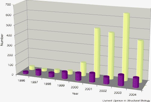
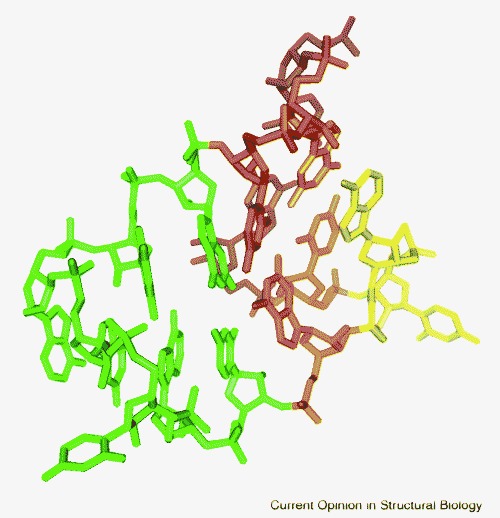
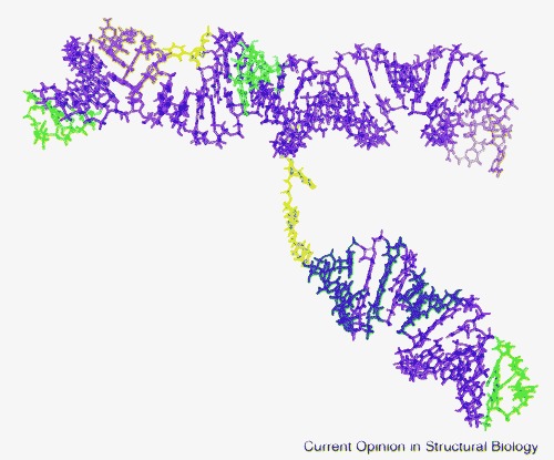
The database of RNA structure has grown tremendously since the crystal structure analyses of ribosomal subunits in 2000-2001. During the past year, the trend toward determining the structure of large, complex biological RNAs has accelerated, with the analysis of three intact group I introns, A- and B-type ribonuclease P RNAs, a riboswitch-substrate complex and other structures. The growing database of RNA structures, coupled with efforts directed at the standardization of nomenclature and classification of motifs, has resulted in the identification and characterization of numerous RNA secondary and tertiary structure motifs. Because a large proportion of RNA structure can now be shown to be composed of these recurring structural motifs, a view of RNA as a modular structure built from a combination of these building blocks and tertiary linkers is beginning to emerge. At the same time, however, more detailed analysis of water, metal, ligand and protein binding to RNA is revealing the effect of these moieties on folding and structure formation. The balance between the views of RNA structure either as strictly a construct of preformed building blocks linked in a limited number of ways or as a flexible polymer assuming a global fold influenced by its environment will be the focus of current and future RNA structural biology.
Figures

References
-
- Ke A., Doudna J.A. Crystallization of RNA and RNA-protein complexes. Methods. 2004;34:408–414. - PubMed
-
- D'Souza V., Dey A., Habib D., Summers M.F. NMR structure of the 101-nucleotide core encapsidation signal of the Moloney murine leukemia virus. J Mol Biol. 2004;337:427–442. - PubMed
-
The RNA structure of the core recognition signal for encapsidation of the positive strand genome of MMLV is reported. This is the largest NMR structure of an RNA determined to date.
-
- Leontis N.B., Westhof E. Analysis of RNA motifs. Curr Opin Struct Biol. 2003;13:300–308. - PubMed
-
- Ban N., Nissen P., Hansen J., Moore P.B., Steitz T.A. The complete atomic structure of the large ribosomal subunit at 2.4 Å resolution. Science. 2000;289:905–920. - PubMed
Publication types
MeSH terms
Substances
Grants and funding
LinkOut - more resources
Full Text Sources
Other Literature Sources
Miscellaneous
