Comparative Study
. 2005 Aug;116(3):276-288.
doi: 10.1016/j.pain.2005.04.014.

Role of NK-1 neurotransmission in opioid-induced hyperalgesia

Affiliations
Comparative Study

Role of NK-1 neurotransmission in opioid-induced hyperalgesia

Tamara King et al. Pain. 2005 Aug.

Abstract

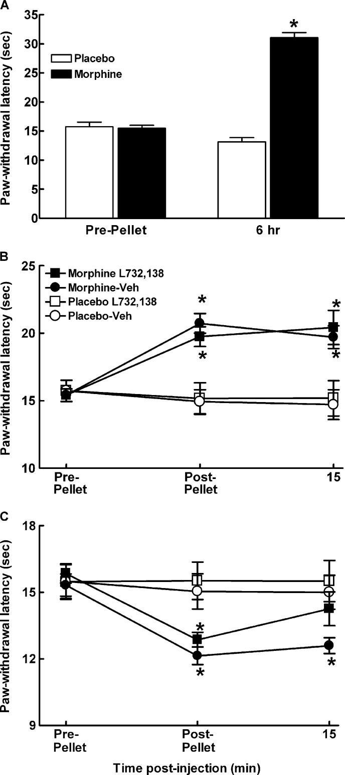
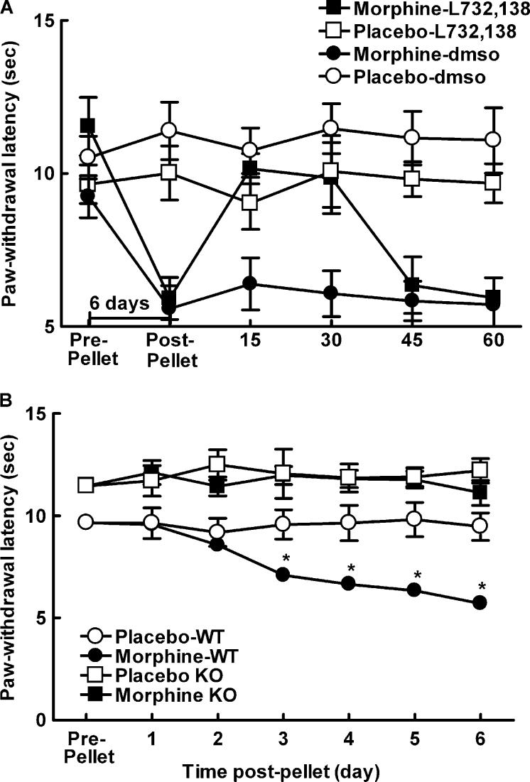
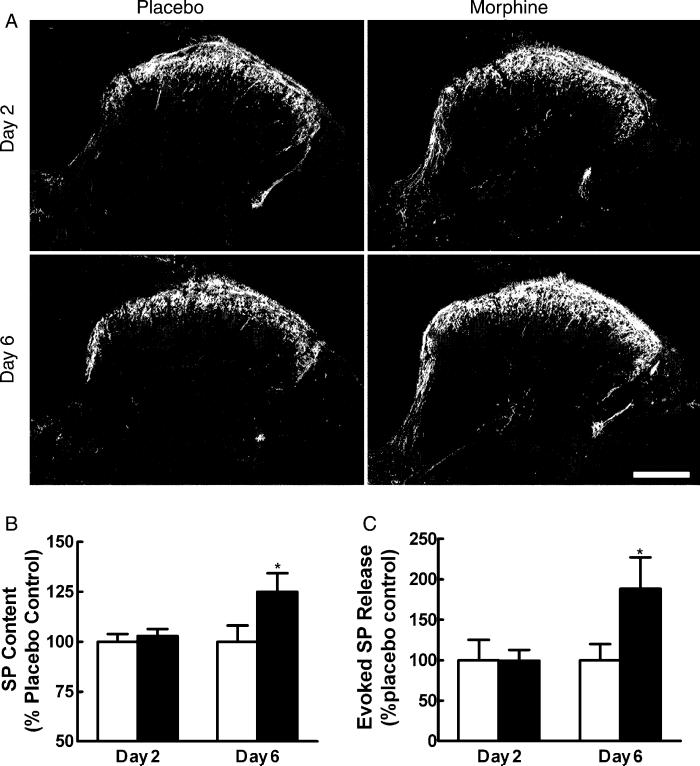
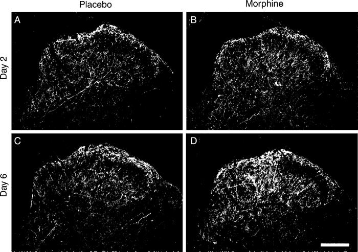
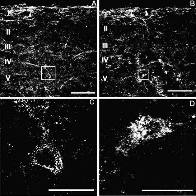
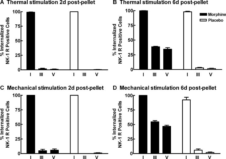
Opiates are among the most important drugs for treatment of moderate to severe pain and prolonged opiate administration is often required to treat chronic pain states. We investigated the neurobiological actions of sustained opiate administration revealing paradoxical pronociceptive adaptations associated with NK-1 receptor function. Sustained morphine delivered over 6 days elicited hyperalgesia in rats and mice during the period of opiate delivery. Sustained morphine administration increased substance P (SP) and NK-1 receptor expression in the spinal dorsal horn. Sustained morphine treatment also enhanced capsaicin-evoked SP release in vitro, and increased internalization of NK-1 receptors in response to noxious stimulation. While NK-1 receptor internalization was observed primarily in the superficial laminae of placebo-treated rats, NK-1 receptor internalization was seen in both superficial and deep lamina of the dorsal horn in morphine-treated animals. Morphine-induced hyperalgesia was reversed by spinal administration of an NK-1 receptor antagonist in rats and mice, and was observed in wildtype (NK-1(+/+)), but not NK-1 receptor knockout (NK-1(-/-)), mice. These data support a critical role for the NK-1 receptor in the expression of sustained morphine-induced hyperalgesia. Additionally, these data indicate that sustained opiate administration induces changes reminiscent of those associated with inflammatory pain. These opiate-induced changes might produce unintended deleterious actions in the course of pain treatment in patients. Understanding of sustained morphine-induced neurochemical changes will help identify approaches that limit the deleterious actions of opiates.


Figures

Fig. 1
Fig. 1
Male Sprague–Dawley rats were implanted with two placebo or morphine pellets. (A) Morphine induced a robust antinociception within 6 h of pellet implant. (B) Rats were tested 2 days post-pellet implantation. Paw-withdrawal latencies of morphine-pelleted animals were longer than placebo-pelleted rats, indicating hypoalgesia, P≤0.05. Intrathecal administration of the NK-1 receptor antagonist, L-732,138 (25 μg/5 μl) or the vehicle control (DMSO) did not alter the responses of placebo or morphine-pelleted rats to the thermal stimulus at 15 min post-injection. (C) Rats were tested 6 days post-pellet implantation. Paw-withdrawal latencies of morphine-pelleted animals were lower than the placebo-pelleted rats, indicating that thermal hyperalgesia emerges by 6 days post-morphine pellet. Paw-withdrawal latencies were measured 15 min after intrathecal administration of the NK-1 receptor antagonist, L-732,138 (25 μg/5 μl). Intrathecal administration of L-732,138 reversed thermal hyperalgesia within 15 min post-administration in morphine-pelleted rats, but did not alter the response of placebo-pelleted animals. Intrathecal administration of the vehicle did not alter responses of the morphine or placebo-pelleted animals. Graphs represent mean±SEM. Each treatment group consisted of 12 rats. * indicates significant difference from pre-pellet baselines.
Fig. 2
Fig. 2
(A) Male C57BL/6×129Sv mice were implanted with one placebo or morphine pellet. All mice showed Straub tail and characteristic stereotypic circling behavior within 30 min of morphine pellet administration. Morphine-treated mice showed decreased paw-withdrawal latencies compared to the placebo-treated animals 6 days following pellet implant, P<0.05, indicating morphine-induced hypersensitivity in these animals. Intrathecal administration of the NK-1 receptor antagonist 6 days post-morphine pellet reversed morphine-induced thermal hypersensitivity within 15 min post-administration, P<0.05, and this reversal lasted approximately 30 min, with paw-flick latencies back to pre-administration baselines 45 min post-administration of L-732,138. Intrathecal administration of L-732,138 did not alter paw-flick latencies of placebo-treated animals. (B) Male NK-1−/− or NK-1+/+ mice were implanted with one placebo or morphine pellet. NK-1−/− mice had longer paw-withdrawal latencies compared to NK-1+/+ mice throughout the testing period, P≤0.05. NK-1+/+ mice treated with morphine showed decreased paw-withdrawal latencies compared to the NK-1+/+ placebo-treated animals starting 3 days post-pellet implant, P≤0.05, indicating morphine-induced hypersensitivity in these animals. Morphine-pelleted NK-1−/− mice showed paw-withdrawal latencies that were equivalent to placebo-pelleted NK-1−/− mice across the entire 6-day testing period, indicating no morphine-induced hypersensitivity in these animals. * indicates significant difference from pre-pellet baselines.
Fig. 3
Fig. 3
(A) Sustained morphine up-regulates SP immunofluorescence in the spinal cord. Tissue from placebo-treated rats is shown in the left column, and that from morphine-treated rats is shown in the right column. No apparent differences in SP immunoreactivity are seen between the spinal sections obtained from placebo-treated and morphine-treated rats 2 days after pellet implant. The staining intensity of SP in tissues from morphine-treated rats on day 6 is enhanced compared to placebo-treated rats on day 6. The enhanced staining is apparent in laminae I and II. Scale bar, 200 μm. (B) Radioimmunoassay shows no differences in SP content between placebo animals in the 2 and 6 days post-pellet implant. Morphine did not alter SP content in rats 2 days post-pellet implant. SP content was elevated 6 days post-morphine pellet implant (*P<0.05). (C) The capsaicin-evoked release of SP from spinal tissues in vitro 2 and 6 days after placebo or morphine pellet implant is represented as the amount of SP above the baseline release for each individual group. Basal levels of SP release did not differ among the treatment groups. Evoked SP release was not different between tissues from placebo- and morphine-treated groups 2 days after pellet implant (P>0.05). However, tissues from the 6-day morphine-treated group showed a significantly greater level of capsaicin-evoked release of SP (*P<0.05). Graphs represent mean±SEM. Each treatment group consisted of 12 rats.
Fig. 4
Fig. 4
Sustained morphine up-regulates spinal NK-1 receptors. Tissue from placebo-treated rats is shown in the left column, and that from morphine-treated rats is shown in the right column. No apparent differences in NK-1 receptor immunoreactivity is seen between the spinal sections obtained from placebo-treated and morphine-treated rats 2 days after pellet implantation (A, B). The staining intensity of NK-1 receptors in tissues from morphine-treated rats on day 6 is enhanced compared to placebo-treated rats on day 6 (C, D). The enhanced staining is apparent in both laminae I/II and in deeper laminae. Scale bar, 200 μm.
Fig. 5
Fig. 5
Sustained morphine-induced NK-1 receptor internalization in deep dorsal horn neurons. Confocal images show that mechanical stimulation (2 min pinch) induced NK-1 receptor internalization in the spinal cord dorsal horn of rats 6 days after placebo (A, C) or morphine (B, D) pellets were implanted subcutaneously. NK-1 receptor internalization after mechanical stimulation is confined to lamina I spinal neurons in placebo-pelleted animals (A). Arrowheads indicate neurons with NK-1 receptor internalization in lamina I spinal neurons. In contrast, sustained exposure to morphine induces NK-1 receptor internalization in deeper laminae III–V neurons as well as in lamina I neurons (B). Arrows indicate lamina I spinal neurons with NK-1 receptor internalization. High power magnification illustrates the difference between deep dorsal horn neurons with and without NK-1 receptor internalization (C, D). Rats that were treated with morphine pellets across 6 days show neurons that have internalized the NK-1 receptor (D). The cytoplasm of these neurons contains bright, immunofluorescent NK-1 receptor-immunoreactive endosomes, defined as an intensely NK-1 receptor-immunoreactive intracellular organelle 0.1–0.7 μm in diameter that are clearly not part of the external plasma membrane (D). In placebo-treated rats, deep dorsal horn neurons contained less than five endosomes per cell (C). In the present study, we considered a cell to have internalized receptors if it contained >20 endosomes. (A) and (C) were produced by superimposition of three optical sections taken at 2.4 μm in sagittal sections of L4, the scale bar is 100 μm; (B) and (D) were produced by superimposition of seven optical sections taken at 0.6 μm at L4, the scale bar is 20 mm.
Fig. 6
Fig. 6
Sustained morphine increases noxious stimulation induced NK-1 receptor internalization in deep dorsal horn neurons. Male Sprague–Dawley rats were implanted with two placebo or morphine pellets. Separate groups were exposed to mechanical (2 min pinch) or thermal (2 min at 52 °C) stimulation 2 and 6 days after pellet implantation; the animals were perfused 5 min post-stimulation, and lumbar spinal cord tissues were collected and processed for NK-1 receptor immunoreactivity. The percentage of NK-1 receptor labeled neurons at the L4 segment that contained internalized NK-1 receptors after thermal or mechanical stimulation were counted in laminae I, III, and V of the spinal cord. The thermal and mechanical stimulation induced internalization of the NK-1 receptor in approximately 100% of the NK-1 receptor labeled lamina I cells across all conditions. (A) and (C) show that under 5% of NK-1 receptor labeled neurons in laminae III–V showed NK-1 receptor internalization after the thermal or mechanical stimulation 2 days after placebo or morphine pellet implantation. (B) and (D) show that 6 days after morphine pellet implants, both the thermal and mechanical stimuli induced an increase in the percentage of NK-1 receptor labeled cells that internalized the NK-1 receptor in laminae III and V compared to the placebo-pelleted animals. Graphs represent mean±SEM. Each treatment group consisted of 3–5 rats.

Similar articles

Cited by

References

    1. Abbadie C, Brown JL, Mantyh PW, Basbaum AI. Spinal cord substance P receptor immunoreactivity increases in both inflammatory and nerve injury models of persistent pain. Neuroscience. 1996;70:201–9. - PubMed
    1. Abbadie C, Trafton J, Liu H, Mantyh PW, Basbaum AI. Inflammation increases the distribution of dorsal horn neurons that internalize the neurokinin-1 receptor in response to noxious and non-noxious stimulation. J Neurosci. 1997;17:8049–60. - PMC - PubMed
    1. Birch PJ, Harrison SM, Hayes AG, Rogers H, Tyers MB. The non-peptide NK-1 receptor antagonist, (+/−)-CP-96,345, produces antinociceptive and anti-oedema effects in the rat. Br J Pharmacol. 1992;105:508–10. - PMC - PubMed
    1. Cahill CM, Coderre TJ. Attenuation of hyperalgesia in a rat model of neuropathic pain after intrathecal pre- or post-treatment with a neurokinin-1 antagonist. Pain. 2002;95:277–85. - PubMed
    1. Cascieri MA, Macleod AM, Underwood D, Shiao LL, Ber E, Sadowski S, Yu H, Merchant KJ, Swain CJ, Strader CD, Fong TM. Characterization of the interaction of N-acyl-L-tryptophan benzyl ester neurokinin antagonists with the human neurokinin-1 receptor. J Biol Chem. 1994;269:6587–91. - PubMed

Publication types

MeSH terms