. 2005 Jul;25(13):5404-16.
doi: 10.1128/MCB.25.13.5404-5416.2005.

Transcription factor NF-kappaB differentially regulates death receptor 5 expression involving histone deacetylase 1

Affiliations

Transcription factor NF-kappaB differentially regulates death receptor 5 expression involving histone deacetylase 1

Shashirekha Shetty et al. Mol Cell Biol. 2005 Jul.

Abstract

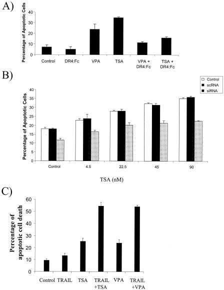
The transcription factor nuclear factor kappaB (NF-kappaB) regulates the expression of both anti-apoptotic and proapoptotic genes. Death receptor 5 (DR5, TRAIL-R2) is a proapoptotic protein considered to be a potential target for cancer therapy, and its expression is mediated by NF-kappaB. The mechanism of NF-kappaB-induced DR5 expression is, however, unknown. Herein, we determined that etoposide-induced DR5 expression requires the first intronic region of the DR5 gene. Mutation of a putative NF-kappaB binding site in this intron eliminates DR5 promoter activity, as do mutations in the p53 binding site in this region. Reduction in p53 expression also blocks p65 binding to the intronic region of the DR5 gene, indicating cooperation between p53 and p65 in DR5 expression. In contrast, the anti-apoptotic stimulus, epidermal growth factor (EGF), fails to increase DR5 expression but effectively activates NF-kappaB and induces p65 binding to the DR5 gene. EGF, however, induces the association of histone deacetylase 1 (HDAC1) with the DR5 gene, whereas etoposide treatment fails to induce this association. Indeed, HDAC inhibitors activate NF-kappaB and p53 and upregulate DR5 expression. Blockage of DR5 activation decreased HDAC inhibitor-induced apoptosis, and a combination of HDAC inhibitors and TRAIL increased apoptosis. This provides a mechanism for regulating NF-kappaB-mediated DR5 expression and could explain the differential roles NF-kappaB plays in regulating apoptosis.


Figures

FIG. 1.
FIG. 1.
The first intronic region of the DR5 gene regulates genotoxin-induced expression. (A) (i) Schematic representation of the human DR5 gene fragment shows the promoter, the exon, and the first intron region containing the two sites that have 99% homology to p53 and NF-κB consensus DNA binding sequence upstream of the luciferase (luc) gene. In contrast, a DR5 promoter alone upstream of the luciferase gene was also constructed. (ii) Putative NF-κB binding sites in the first intronic region of DR5 in mice and rats compared to that of humans. (B) MCF-7 cells were transfected with promoter alone (black bars) or promoter-intron luciferase reporter plasmid (white bars) along with a β-Gal reporter plasmid as a control. Cells were treated with etoposide (100 μM) over a 6-h time course. DR5 promoter activity is represented by the increase in luciferase activity normalized to β-Gal expression. Error bars represent the standard deviations for three independent experiments. (C) MCF-7 cells were also transfected with promoter alone (white bars) or promoter-intron luciferase reporter plasmid (black bars) and treated with EGF (1 μg/ml) over a 6-h time course. The cells were lysed and a luciferase assay was performed as described above.
FIG. 2.
FIG. 2.
DR5 transcriptional activity requires NF-κB activation. (A) (i) HEK293 cells expressing vector alone and ΔIκB were transfected with a luciferase reporter plasmid containing DR5 promoter-intron region and a β-Gal reporter plasmid (transfection control). The cells were harvested after 24 h and lysed. A luciferase assay was performed as described in Materials and Methods. Relative luciferase units represent DR5 promoter activity. Error bars represent the standard deviations for three independent experiments. (ii) HEK293 cells were transiently transfected with vector alone (lane 1) or ΔIκB along with a luciferase reporter plasmid containing DR5 promoter-intron region and a β-GAL reporter plasmid (transfection control; lane 2). The cells were then treated with 100 μM etoposide for 4 h and analyzed as described above. The cells were also lysed and Western blotted for IκB and reprobed for actin (loading control). (iii) HEK293 cells were transfected with a BAX promoter luciferase construct with empty vector (control) or ΔIκB. The cells were treated with 100 μM etoposide for 16 h, and luciferase activity was measured as described above. (B) The NF-κB putative binding site was mutated at GGGAGTATCGC, where nucleotides underlined represent point mutations. HEK293 and MCF-7 cells were transfected with a luciferase reporter plasmid alone (Vector), containing the wild type DR5 promoter-intron region (Promoter), or containing a mutation in the putative NF-κB binding site of the DR5 intron region (Promoter NFκB mutation). β-Gal reporter plasmid was used as a control for transfection efficiency. The cells were harvested after 24 h and lysed, and a luciferase assay performed as described in Materials and Methods. Relative luciferase units represent DR5 promoter activity. Error bars represent the standard deviations for three independent experiments. (C) Cells were transfected with wild-type or p65 mutant constructs as described above. A time course of 16 h following etoposide treatment was performed, and the increase in luciferase activity was determined. Standard errors represent three independent experiments.
FIG. 3.
FIG. 3.
Overexpression of NF-κB subunit p65 induces DR5 transcriptional activity, and NF-κB binds to the DR5 gene in the first intron. (A) (i) HEK293 and MCF-7 cells were cotransfected with vector alone (lanes C), p65 cDNA containing CMV4 expression vector (lane p65) or p50 cDNA-containing pEV expression vector (lane p50) with a DR5 promoter-intron region luciferase reporter plasmid and a β-Gal reporter plasmid as a transfection control. HEK293 cells were lysed and Western blotted for p50 or p65 and reprobed with actin antibodies (loading control). (ii) Cells were harvested after 24 h and lysed, and a luciferase assay was performed as described in Materials and Methods. DR5 promoter activity is represented by the increase in luciferase activity. Error bars represent the standard deviations for three independent experiments. (iii) HEK293 cells were transiently transfected with vector alone or p65 in combination with the DR5 promoter (Wt), promoter with the p53 binding site mutated (p53 mut), or promoter with the NF-κB binding site mutated (NFκB mut). The cells were then treated with etoposide for 6 h and analyzed as described above. (B) HEK293 cells stably expressing p65 or p50 subunits were lysed and analyzed by Western blotting for DR5 protein expression as described in Materials and Methods. HEK293 cells stably expressing vector alone (Vector) were used a control. β-Actin was used as a loading control. (C) Electromobility shift assay using the NF-κB binding site in the intronic region-specific probe following treatment with etoposide (100 μM) over a 6-h time course was performed with HEK293 cells. Where indicated, nuclear extracts from cells treated with etoposide for 4 h were mixed with anti-p65 prior to incubation with the NF-κB intronic probes. The lower band represents p50 homodimers. (D) HEK293 cells untreated or treated with etoposide (100 μM) were used in a ChIP assay with anti-p65 antibodies. Sample with no antibody added was used as a negative control, whereas genomic DNA was used as the input control. Precipitated DNA was analyzed by PCR using primers specific for (i) the DR5 exon 1 and intron 1 region or (ii) an upstream region of the DR5 gene (1,353 to 1,832 nucleotides). All experiments were repeated three times. (E) 3T3 murine fibroblast cells, parental or lacking p65 expression, were transfected with the DR5 promoter-intron region luciferase reporter plasmid and a β-Gal reporter plasmid. The cells were then treated with 100 μM etoposide and analyzed as described above. Wt, wild type.
FIG. 4.
FIG. 4.
The tumor suppressor p53 is also required for DR5 promoter activity. (A) The p53 site was mutated at GGGAATATCCGGGCAAGACG, where underlined nucleotides represent point mutations. HEK293 and MCF-7 cells were transfected with luciferase reporter plasmid alone (Vector), containing wild-type DR5 promoter-intron region (Promoter), or containing mutations in the p53 binding site in the DR5 promoter-intron region (Promoter p53 mutation). β-Gal reporter plasmid was used as a control for transfection efficiency. The cells were harvested after 24 h and lysed, and a luciferase assay was performed as described in Materials and Methods. Relative luciferase units represent DR5 promoter activity. Error bars represent the standard deviations for three independent experiments. (B) HEK293 cells untreated and treated with etoposide (100 μM) were used in a ChIP assay with anti-p53 antibody. Sample with no antibody added was used as the negative control, whereas genomic DNA was used as the input control. Immunoprecipitated DNA was analyzed by PCR using primers specific for DR5 exon 1 and intron 1 regions. (C) HEK293 cells were transfected with siRNA against p53 or scrambled siRNA followed by etoposide (100 μM) treatment. The cells were lysed, and a p53 or p65 ChIP assay was preformed as described above.
FIG. 5.
FIG. 5.
EGF fails to increase DR5 expression but induces NF-κB activation and binding to the DR5 gene. (A) MCF-7 cells were treated with etoposide (100 μM) for the indicated times and then lysed. The RNase protection assay (RPA) was performed by using an hAPO3d probe set (Pharmingen) as described in Materials and Methods. Error bars represent the standard deviations for three independent experiments. The cells untreated or treated with etoposide (24 h) were also Western blotted for DR5 and actin (loading control). (B) MCF-7 cells were treated with EGF (1 μg/ml) for the times indicated. The RPA was performed as described above. Error bars represent the standard deviations for three independent experiments. The cells untreated or treated with EGF (24 h) were also Western blotted for DR5 and actin (loading control). (C) An electromobility shift assay using the NF-κB binding site in the intronic region-specific probe was performed with HEK293 cells following treatment with EGF (1 mg/ml) over a 120-min time course. Where indicated, nuclear extracts from cells stimulated with EGF for 30 min were mixed with anti-p65 or anti-p50 antibodies prior to incubation with NF-κB intronic probes. (D) HEK293 cells untreated or treated with EGF (1 μg/ml) over a 4-h time course were used in a ChIP assay with anti-p65 antibodies. Isotype-matched antibodies were used as the negative control, whereas genomic DNA was used as the input control. Precipitated DNA was analyzed by PCR using primers specific for the DR5 exon 1 and intron 1 region. (E) A ChIP assay was also performed with anti-p53 antibodies on HEK293 cells treated with EGF as described above. (F) HEK293 cells were treated with etoposide (100 μM) or EGF (1 μg/ml) over the indicated times. The cells were then lysed and Western blotted for p53 and actin (loading control).
FIG. 6.
FIG. 6.
DR5 gene is acetylated following etoposide but not EGF treatment. HEK293 cells were untreated, treated with etoposide (100 μM) over a 16-h time course (A), or treated with EGF (1 μg/ml) over an 8-h time course (B). These cells were used for a ChIP assay with anti-acetyl histone H3. Isotype matched antibody was used as a negative control. Precipitated DNA was analyzed by PCR using primers specific for DR5 exon 1 and intron 1 regions. As a positive control, histone acetylation of the constitutively expressing pS2 gene was used as previously published (43a).
FIG. 7.
FIG. 7.
HDAC1 is recruited to the DR5 gene following EGF but not etoposide treatment mediated by NF-κB. (A) HEK293 cells that were untreated, treated with etoposide (100 μM), or treated with EGF (1 μg/ml) over an 8-h time course were used in a ChIP assay with anti-HDAC1 antibody. Sample with no antibody added was used as the negative control. Genomic DNA was used as the input control. (B) HEK293 cells were treated with etoposide (100 μM) or EGF (1 μg/ml) over the indicated time course. The cells were lysed, immunoprecipitated with anti-p65 antibodies, and Western blotted for HDAC1. The blots were stripped and reprobed for p65 for equal loading. (C) HEK293 cells with vector alone or expressing ΔIκB wereuntreated or treated with EGF (1 μg/ml) over a 4-h time course. These cells were used in a ChIP assay with anti-HDAC1 antibodies as previously described. (D) HEK293 cells were treated with EGF (1 μg/ml) over a 2-h time course. The cells were then lysed and immunoprecipitated with anti-HDAC1 or anti-p65 antibodies as described in Materials and Methods for ChIP assays. Lysates from the p65 immunoprecipitation were reimmunoprecipitated with anti-HDAC1 antibodies. The immunoprecipitated DNA was isolated, and PCR was performed to detect the first intron region of the DR5 gene. Input represents the input DNA from the cell lysates, and the negative control (-ve Control) represents sample with no antibody control.
FIG. 8.
FIG. 8.
Inhibition of HDAC activity increases DR5 expression. (A) (i) HEK293 cells were untreated (C) or treated with TSA (45 nM), VPA (500 μg/ml), or etoposide (Eto; 100 μM) for 24 h. An RNase protection assay was performed by using the hAPO3d probe set (Pharmingen) as described in Materials and Methods. A similar trend was observed in three different experiments. (ii) The amount of DR5 mRNA was quantified by a phosphorimager as the fold increase normalized to loading control (L32). (B) HEK293 cells were untreated, treated with EGF (1 μg/ml) alone or TSA (45 nM) alone or EGF and TSA in combination for 24 h. The cells were lysed and Western blotted with antibodies against DR5 or DR4. The blots were stripped and reprobed with antibodies against actin (loading control). (C) Cells were transfected with luciferase reporter gene constructs containing a promoter with NF-κB binding sites (NFκB-Luc) or a promoter with p53 binding sites (p53-Luc). The cells were then treated with TSA (45 nM) for 16 h and the increase in luciferase activity was determined. (D) The cells were also treated with TSA (45 nM) for 16 h and lysed. A ChIP (i.p.) assay was performed using antibodies against p65, p53, or HDAC1 as described in Materials and Methods. Sample with no antibody added was used as a negative control; genomic DNA was used as the input control.
FIG. 9.
FIG. 9.
HDAC inhibitor-induced apoptosis involves DR5 activation. (A) MCF-7 cells were treated with TSA (45 nM) or VPA (500 μg/ml). The cells were also incubated with DR4:Fc protein as indicated. The amount of apoptosis was determined by acridine orange. Error bars represent the standard errors for three independent experiments. (B) HEK293 cells were transfected with siRNA against DR5 or scrambled RNA (scRNA). The cells were then treated with a range of concentrations of TSA, as indicated, for 48 h. The amount of apoptosis was determined by acridine orange staining. (C) MCF-7 cells were treated with TSA (45 nM), VPA (500 μg/ml), TRAIL (100 ng/ml) individually or in combination, as indicated, for 36 h. The percentages of apoptotic cells were determined by staining the cells with acridine orange to detect condensed DNA. Apoptotic cells were scored, with a total of 300 cells being counted for each condition. Standard deviations were determined on the basis of three independent experiments.

References

    1. Aron, J. L., M. R. Parthun, G. Marcucci, S. Kitada, A. P. Mone, M. E. Davis, T. Shen, T. Murphy, J. Wickham, C. Kanakry, D. M. Lucas, J. C. Reed, M. R. Grever, and J. C. Byrd. 2003. Depsipeptide (FR901228) induces histone acetylation and inhibition of histone deacetylase in chronic lymphocytic leukemia cells concurrent with activation of caspase 8-mediated apoptosis and down-regulation of c-FLIP protein. Blood 102:652-658. - PubMed
    1. Ashburner, B. P., S. D. Westerheide, and A. S. Baldwin, Jr. 2001. The p65 (RelA) subunit of NF-κB interacts with the histone deacetylase (HDAC) corepressors HDAC1 and HDAC2 to negatively regulate gene expression. Mol. Cell. Biol. 21:7065-7077. - PMC - PubMed
    1. Ashkenazi, A., and V. M. Dixit. 1998. Death receptors: signaling and modulation. Science 281:1305-1308. - PubMed
    1. Baldwin, A. S. 2001. Control of oncogenesis and cancer therapy resistance by the transcription factor NF-κB. J. Clin. Investig. 107:241-246. - PMC - PubMed
    1. Baldwin, A. S., Jr. 1996. The NF-κB and IκB proteins: new discoveries and insights. Annu. Rev. Immunol. 14:649-683. - PubMed

Publication types

MeSH terms