. 2005 Jul;25(13):5552-66.
doi: 10.1128/MCB.25.13.5552-5566.2005.

Histone acetyltransferase activity of p300 is required for transcriptional repression by the promyelocytic leukemia zinc finger protein

Affiliations

Histone acetyltransferase activity of p300 is required for transcriptional repression by the promyelocytic leukemia zinc finger protein

Fabien Guidez et al. Mol Cell Biol. 2005 Jul.

Abstract

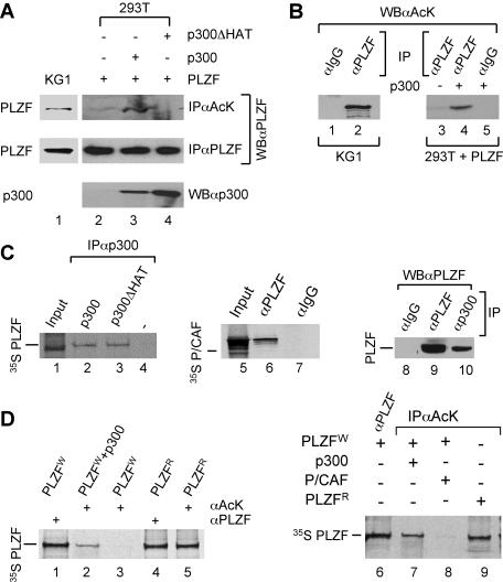
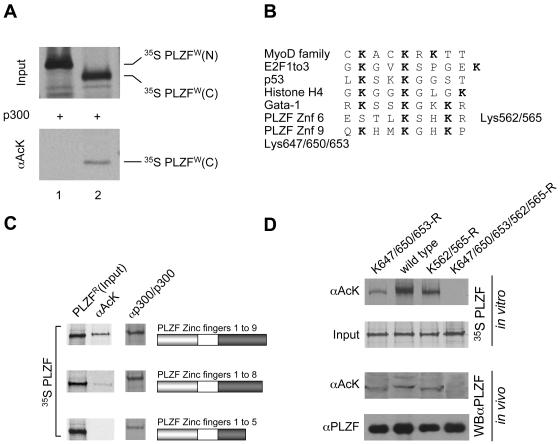
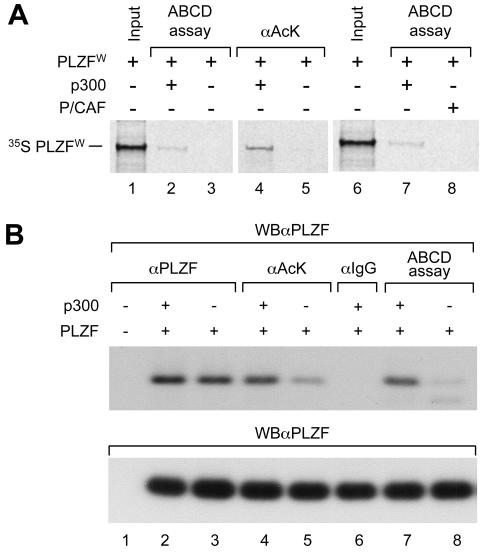
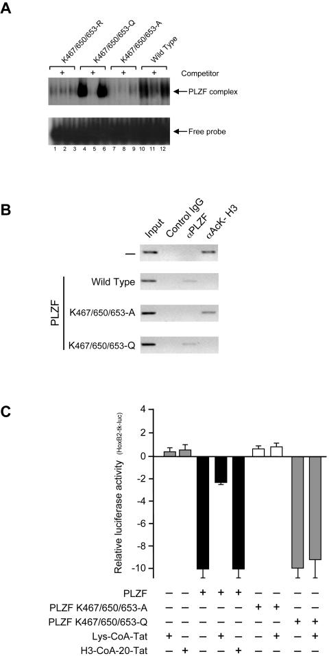
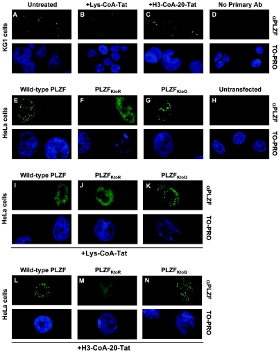
Histone acetyltransferase (HAT) activities of proteins such as p300, CBP, and P/CAF play important roles in activation of gene expression. We now show that the HAT activity of p300 can also be required for down-regulation of transcription by a DNA binding repressor protein. Promyelocytic leukemia zinc finger (PLZF), originally identified as a fusion with retinoic acid receptor alpha in rare cases of all-trans-retinoic acid-resistant acute promyelocytic leukemia, is a transcriptional repressor that recruits histone deacetylase-containing corepressor complexes to specific DNA binding sites. PLZF associates with p300 in vivo, and its ability to repress transcription is specifically dependent on HAT activity of p300 and acetylation of lysines in its C-terminal C2-H2 zinc finger motif. An acetylation site mutant of PLZF does not repress transcription and is functionally deficient in a colony suppression assay despite retaining its abilities to interact with corepressor/histone deacetylase complexes. This is due to the fact that acetylation of PLZF activates its ability to bind specific DNA sequences both in vitro and in vivo. Taken together, our results indicate that a histone deacetylase-dependent transcriptional repressor can be positively regulated through acetylation and point to an unexpected role of a coactivator protein in transcriptional repression.


Figures

FIG. 1.
FIG. 1.
PLZF protein is acetylated by p300 in vivo. (A) To assess PLZF acetylation, PLZF was immunoprecipitated with a rabbit polyclonal anti-acetyl-lysine antibody (αAcK) prior to Western blotting with the monoclonal anti-PLZF antibody (WBαPLZF). Endogenous PLZF in KG1 cells (lane 1); PLZF expressed in transfected 293T cells in the absence of p300 (lane 2) or in the presence of wild-type (lane 3) or the ΔHAT mutant (lane 4) of p300. To assess the level of expression of p300 proteins in each experiment, Western analysis was performed with a polyclonal anti-p300 antibody (WBαp300). Results of protein expression are shown in lanes 3 and 4 for wild-type p300 and p300ΔHAT, respectively. (B) Similar results reflecting the presence of endogenously acetylated PLZF were obtained using anti-PLZF and anti-acetyl-lysine antibodies for immunoprecipitation and Western blotting, respectively. Immunoprecipitation using anti-Gal4 antibody was used as a control (lanes 1 and 5). Acetylation of PLZF was examined in KG1 (lanes 1 and 2) and transfected 293T cells (lanes 3 to 5) as indicated. (C) PLZF interacts with p300 in vivo and in vitro. In vitro 35S-labeled PLZF was translated using the rabbit reticulocyte system (see input lane 1) and incubated with unlabeled p300 (lane 2) and p300ΔHAT (lane 3) proteins or unprogrammed rabbit reticulocyte (lane 4). Anti-p300 antibody (αp300) was used for coimmunoprecipitation to evaluate interactions between the PLZF and p300 proteins. Similarly, 35S-labeled P/CAF was coimmunoprecipitated with PLZF (lane 6) using anti-PLZF antibody (αPLZF) but not anti-Gal4 control (αIgG). Whole-cell extracts from KG1 were subjected to immunoprecipitation with anti-p300 (αp300, lane 10), polyclonal anti-PLZF (αPLZF, lane 9), or an irrelevant anti-Gal4 IgG (αIgG, lane 8) followed by immunoblotting with a monoclonal anti-PLZF antibody (WBαPLZF). (D) In vitro acetylation of PLZF by p300 proteins. In vitro 35S-labeled PLZF was translated using the wheat germ (W) or rabbit reticulocyte (R) system. The acetylation level of PLZF was assessed by immunoprecipitation of 35S-labeled PLZF with an anti-acetyl-lysine antibody (lanes 2, 3, 5, and 7 to 9) followed by SDS gel electrophoresis and autoradiography of the dried gel. To assess the amount of PLZF in a given sample, immunoprecipitations were also performed with an anti-PLZF antibody (lanes 1, 4, and 6). In vitro-translated PLZF (using wheat germ lysate) was incubated with acetyl-CoA and unprogrammed reticulocyte lysate (PLZFw, lane 3) or in vitro-translated p300 (PLZFw+p300, lanes 2 and 7) or P/CAF (lane 8) for 1.5 h at 30°C and assessed for acetylation.
FIG. 2.
FIG. 2.
Inhibition of p300 reduces PLZF acetylation in vivo. (A) Whole-cell lysates were prepared from KG1 cells untreated (lane 1) or treated overnight with the specific inhibitor of p300, Lys-CoA-Tat 10 μM (lane 2), or P/CAF, H3-CoA-20-Tat 10 μM (lane 3). Acetylated PLZF and total protein were immunoprecipitated using the anti-acetyl lysine (αAcK) or anti-PLZF (αPLZF) polyclonal antibodies, and immunoprecipitates were analyzed by Western blotting with a monoclonal antibody raised against PLZF (WBαPLZF). Enhanced chemiluminescence was used for detection (both panels). (B and C) Specific inhibition of p300 (B) and P/CAF (C) activities by Lys-CoA-Tat and H3-CoA-20-Tat in KG1 cells was controlled using a luciferase-based reporter with Gal4 binding sites upstream of the herpes simplex virus thymidine kinase minimal promoter and cotransfected Gal4DBD-p300 (B) or Gal4DBD-P/CAF (C) expression vector.
FIG. 3.
FIG. 3.
PLZF is acetylated in the zinc finger DNA binding region. (A) N- and C-terminal parts of PLZF (amino acids 1 to 455 and 455 to 673, respectively) were expressed and labeled in vitro with [35S]methionine using the wheat germ translation system. In vitro-translated proteins were acetylated in the presence of acetyl-CoA and p300. The degree of acetylation was evaluated by immunoprecipitation with anti-acetyl lysine (αAcK) antibodies (lower panel). (B) Alignment of previously described acetylation sites in other proteins and the potential PLZF sites. (C) Mapping of PLZF acetylation sites. Deletion mutants of PLZF were translated in vitro using rabbit reticulocyte lysate and [35S]methionine and were tested for acetylation (PLZFR) by immunoprecipitation with anti-acetyl-lysine antibodies (αAcK). Corresponding deletion mutants are represented by schematics in which the grey box represents the POZ domain of PLZF and the dark box represents its zinc finger region. The input of each PLZF protein is shown in the column labeled PLZFR. Interactions of PLZF proteins with p300 were tested in vitro by immunoprecipitation using p300 protein immobilized on a protein A/G matrix with a p300 polyclonal antibody (αp300/p300) as bait. (D) PLZF lysine mutants assessed for acetylation in vitro and in vivo. Lysine (K) residues of interest were changed to arginines (R). A mutant lacking the zinc finger 9 acetylation site (K647/650/653-R), a mutant lacking the zinc finger 6 acetylation site (K562/565-R), and the double mutant (K647/650/653/562/565-R) were compared to wild-type PLZF (wild type). In vitro and in vivo experiments were performed with in vitro 35S-labeled PLZF proteins and transfected 293T cell extracts, respectively. The acetylation level of PLZF was assessed by immunoprecipitation of 35S-labeled PLZF with an anti-acetyl-lysine antibody (αAcK). Polyclonal anti-PLZF antibody (αPLZF) was used to immunoprecipitate the total protein (input). For in vivo analysis, proteins were immunoprecipitated as above, and immunoprecipitates were subjected to Western blot analysis with monoclonal anti-PLZF antibodies (WBαPLZF). Enhanced chemiluminescence was used for detection.
FIG. 4.
FIG. 4.
Repression activity of PLZF is regulated by p300 HAT activity. The repression activity of PLZF mutant K647/650/653-R, deficient in acetylation of zinc finger 9 lysines (PLZFmut), was tested in transfection experiments using the luciferase reporter with a herpes simplex virus thymidine kinase promoter and with two upstream PLZF binding sites as identified in the HoxB2 r3/r5-enhancer (38). Expression vectors for PLZF and PLZFmut (A) and their respective GAL4 fusions (B) were cotransfected with the indicated reporter vectors and 50 ng of CMV-lacZ plasmid as a control for transfection efficiency. Where indicated, the expression vector for either p300 or p300ΔHAT was also cotransfected. The activity of the PLZF proteins and GAL4-PLZF fusions were assayed by chromatin immunoprecipitation to determine the level of acetylated histone H4 in the proximity of the PLZF or GAL4 binding and by assaying for luciferase activity; 10% of each cell lysate was used to determine the amount of DNA prior to the immunoprecipitation (input DNA). Levels of transiently expressed proteins were monitored by Western blot analysis using an anti-PLZF or an anti-GAL4(DBD) antibody.
FIG. 5.
FIG. 5.
PLZF lysine mutants assessed for transcriptional activity. Lysine (K) residues in the ninth zinc finger of PLZF were changed to arginines (R) by introducing single point mutation in the PLZF DNA coding sequence. A mutant lacking all the zinc finger 9 acetylation sites (K647/650/653-R) and each mutation separately (K647-R, K650-R or K653-R), were compared to wild-type PLZF (wild type) in its ability to repress transcription using cotransfection experiments in 293T cells. For each mutant, equal amounts of each expression vector and luciferase reporter with a minimal herpes simplex virus thymidine kinase promoter and two PLZF binding sites were cotransfected into 293T cells, and luciferase activity was determined 24 h later. The expression vector for the p300 HAT was cotransfected in each case to enhance PLZF-mediated repression of the luciferase reporter. Each lysine mutation occurring in zinc finger 9 has an impact on the repression activity of the PLZF protein. Levels of transiently expressed proteins were monitored by Western blot analysis using an anti-PLZF antibody (WBαPLZF).
FIG. 6.
FIG. 6.
Overexpression of p300 stimulates acetylation of PLZF and its ability to bind DNA. (A) In vitro 35S-labeled PLZF was translated using the wheat germ system ([35S] PLZFW). In vitro-translated PLZF was incubated with acetyl-CoA and in vitro-translated p300 (lanes 2, 4, and 7) or in vitro-translated P/CAF (lane 8) or an equal amount of unprogrammed reticulocyte lysate (lanes 3 and 5) for 1.5 h at 30°C. Levels of PLZF acetylation and PLZF DNA affinity were assessed by immunoprecipitation with an anti-acetyl lysine antibody (αAcK, lanes 4 and 5) or DNA binding assay (ABCD assay, lanes 2, 3, 7, and 8). Samples were electrophoresed, and the dried gel was autoradiographed. (B) PLZF was expressed in 293T cells after transfection in the absence or presence of p300 overexpression. Total PLZF present in each sample was quantified by Western blot analysis (WBαPLZF, lower panel). PLZF-containing cell extracts were subjected to immunoprecipitation with a control antibody (αIgG) an anti-PLZF antibody (αPLZF) or an anti-acetyl lysine antibody (αAcK). The ability of PLZF to bind DNA was assessed using the ABCD binding assay. As shown in lanes 2 and 3, coexpression of p300 does not affect the level of expression of PLZF. However, the level of PLZF acetylation (lane 4 versus lane 5) and the affinity of PLZF for DNA (lane 7 versus lane 8) are sharply increased with overexpression of p300.
FIG. 7.
FIG. 7.
Acetylated lysine residues are critical for PLZF DNA binding and trans-repressing activities. (A) EMSA shows binding of wild-type (lanes 10 to 12) and mutant PLZF proteins expressed transiently in 293T cells (K-to-A in lanes 7 to 9, K-to-Q in lanes 4 to 6 and K-to-R in lanes 1 to 3) to 32P-end-labeled double-stranded oligonucleotide containing three PLZF binding sites. For the competition assays, a 200-fold molar excess of unlabeled oligonucleotides (lanes 2, 5, 8, and 11) was included in the binding reaction. (B) Expression vectors for wild-type PLZF and the K-to-A and K-to-Q mutants were transiently transfected in 293T cells together with a reporter vector containing PLZF binding sites. Cross-linked chromatin was subjected to immunoprecipitation with an anti-PLZF antibody (αPLZF), an anti-acetylated histone H3 (αAck-H3), and an irrelevant anti-GAL4(DBD) (control IgG) antibodies. Coimmunoprecipitated DNA was analyzed by semiquantitative PCR using primers flanking the PLZF binding sites. The PCR products were electrophoresed on an agarose gel (inverted image shown); 10% of each cell lysate was used to determine the amount of DNA prior to the immunoprecipitation (input). Levels of protein expression from transfected vectors were monitored byWestern blotting using an anti-PLZF antibody (data not shown). (C) Expression vectors for PLZF and the indicated PLZF mutants were cotransfected with a reporter containing PLZF binding sites (HoxB2-tk-luc) and 50 ng of CMV-lacZ plasmid as a control for transfection efficiency. Where indicated, Lys-CoA-Tat (10 μM) and H3-CoA-20-Tat (10 μM) treatment was applied overnight. The levels of expression of transiently expressed proteins were monitored by Western blot analysis using an anti-PLZF antibody (not shown). In experiments leading to the results shown in panels B and C, the expression vector for p300 was cotransfected to enhance and maximize PLZF activity.
FIG. 8.
FIG. 8.
Acetylated PLZF localizes in specific subnuclear compartments. The nuclear localization pattern of PLZF (and indicated mutants) was analyzed in KG1 (A to D) and transfected HeLa (E to N) cells by indirect immunofluorescence and confocal microscopy. As reported previously (43), punctate nuclear distribution of endogenous PLZF was observed in KG1 cells (A). However, only diffuse nuclear localization was observed when cells were incubated overnight with 10 μM of Lys-CoA-Tat (B), whereas 10 μM of P/CAF inhibitor H3-CoA-20-Tat had no effect on the punctate localization pattern of the PLZF protein (C). No immunofluorescence signal was observed when primary anti-PLZF monoclonal antibody was omitted from the experimental procedure (D). Both the wild-type (E) and K-to-Q mutant (G) of PLZF but not the zinc finger 9 acetylation-deficient K-to-R mutant (F), localized to a speckled nuclear pattern when transiently expressed in HeLa cells. As HeLa cells do not express PLZF, no staining was observed in untransfected controls (H). Consistent with other results, treatment of transfected HeLa cells with the p300 inhibitor Lys-CoA-Tat led to a loss of speckled nuclear localization for wild-type PLZF (I) but not for the K-to-Q mutant (K), which mimics PLZF that is constitutively acetylated in the ninth zinc finger. As before, P/CAF inhibitor had no effect on the nuclear localization of transiently expressed PLZF proteins (L to N). In all cases HeLa cells were transfected with 3 μg of a given expression vector per well in six-well plates, allowed to grow for 48 h, and then treated overnight with 10 μM of p300 and P/CAF inhibitors as indicated.
FIG. 9.
FIG. 9.
PLZF K-to-A fails to inhibit SAOS cell growth. (A and B) At 24 h after transfection, the cells were diluted 1:40 and incubated in Dulbecco's modified Eagle's medium supplemented with 10% fetal calf serum and 0.5 mg/ml G418 for 3 weeks. After staining with Giemsa, the colonies grown in three plates were counted. Panel B shows the number of colonies counted on the plates transfected with wild-type PLZF (grey). The empty vector was used as a control (black). (B) The number of colonies grown in the presence of PLZF with lysines in the ninth zinc finger mutated to alanines (K-to-A) (grey) compared with the empty vector (pcDNA3.1[-]) (black) is shown. Statistical significance was determined by a two-tailed Student's t test. (C) At 24 h after transfection, cells expressing wild-type PLZF (Pwt) or PLZF with lysines in the ninth zinc finger mutated to alanines (PKtoA) were lysed with 1% NP-40 and the resulting lysates were blotted with antibodies directed against PLZF or glyceraldehyde-3-phosphate dehydrogenase (GAPDH) (upper and lower panels, respectively). The expression levels of wild-type PLZF and the PLZF K-to-A mutant were equivalent.
FIG. 10.
FIG. 10.
Code-based prediction of PLZF zinc finger-DNA interactions (17, 74, 75). Zinc fingers are shown aligned with experimental and code-predicted DNA binding sites (boxed). The zinc ion in each finger is highlighted in red. Arrows indicate juxtapositioning between individual amino acids (in the binding helices of the zinc fingers) and DNA bases. Amino acids are denoted by the single-letter code, and heterogeneous DNA bases are written with the universal base degeneracy code. Darker blue shading of individual DNA base boxes denotes a stronger match between the predicted and experimental sequences. Two putative models of DNA-binding are proposed. (A) All nine fingers of PLZF are assumed to be canonical in order to obtain a crude prediction of the overall binding preference. Fingers 2 to 4 and 7 to 9 have the best matches with the minimal DNA subsite 5′-TACTGTAC-3′ (hatched underlining). (B) In a refined prediction, fingers 1 to 4 are discarded as they contribute minimally to binding in EMSA (38) (data not shown). Note that fingers 6 to 8 are linked by canonical TGEKP-type linkers, commonly found in DNA-binding zinc fingers. Accordingly, fingers 6 to 8 give the best fit with the minimal DNA subsite 5′-TACTGTAC-3′ (hatched underlining). The C-terminal acetylated lysine residues (K-Ac; pink) are shown contacting DNA, but it is unlikely that such DNA contacts alone could account for the binding transition observed between unmodified and acetylated PLZF. (C) Models of PLZF homodimerization through the N-terminal POZ/BTB domain (2). Dotted arrows indicate potential dimerization. Applying the binding model in panel B, PLZF dimers might bind to two separate, almost palindromic DNA subsites (dark blue). Note that alternative dimer orientations are also possible by binding to the antiparallel DNA strands.

Similar articles

Cited by

References

    1. Aalfs, J. D., and R. E. Kingston. 2000. What does ‘chromatin remodeling’ mean? Trends Biochem. Sci. 25:548-555. - PubMed
    1. Ahmad, K. F., C. K. Engel, and G. G. Prive. 1998. Crystal structure of the BTB domain from PLZF. Proc. Natl. Acad. Sci. USA 95:12123-12128. - PMC - PubMed
    1. Ahmad, K. F., A. Melnick, S. Lax, D. Bouchard, J. Liu, C. L. Kiang, S. Mayer, S. Takahashi, J. D. Licht, and G. G. Prive. 2003. Mechanism of SMRT corepressor recruitment by the BCL6 BTB domain. Mol. Cell 12:1551-1564. - PubMed
    1. Ball, H. J., A. Melnick, R. Shaknovich, R. A. Kohanski, and J. D. Licht. 1999. The promyelocytic leukemia zinc finger (PLZF) protein binds DNA in a high molecular weight complex associated with cdc2 kinase. Nucleic Acids Res. 27:4106-4113. - PMC - PubMed
    1. Bannister, A. J., E. A. Miska, D. Gorlich, and T. Kouzarides. 2000. Acetylation of importin-alpha nuclear import factors by CBP/p300. Curr. Biol. 10:467-470. - PubMed

Publication types

MeSH terms