Comparative Study
. 2005 Jul;25(13):5616-25.
doi: 10.1128/MCB.25.13.5616-5625.2005.

Lack of the central nervous system- and neural crest-expressed forkhead gene Foxs1 affects motor function and body weight

Affiliations
Comparative Study

Lack of the central nervous system- and neural crest-expressed forkhead gene Foxs1 affects motor function and body weight

Mikael Heglind et al. Mol Cell Biol. 2005 Jul.

Abstract

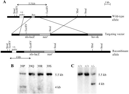
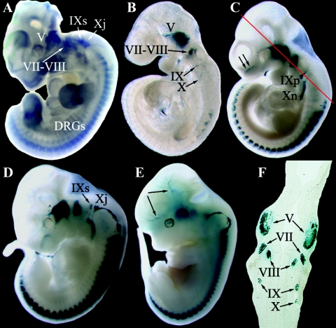
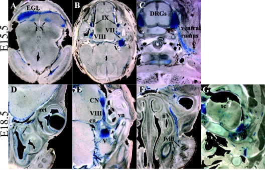
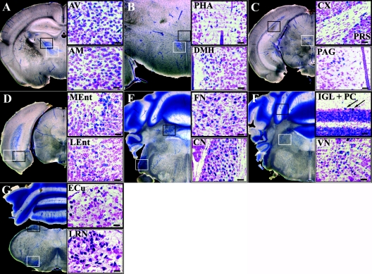
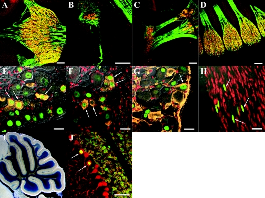
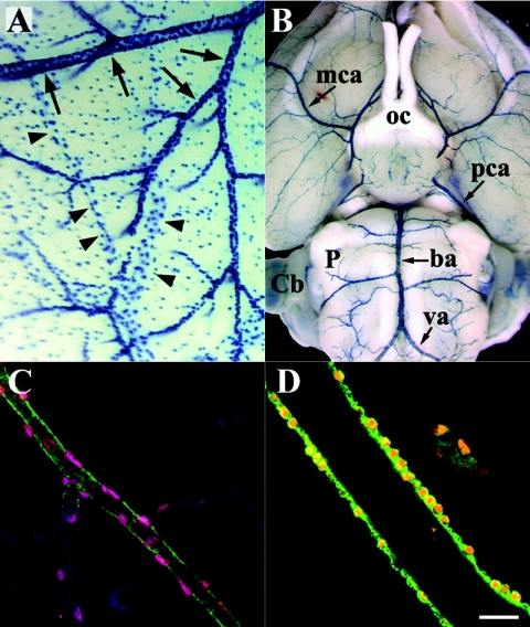
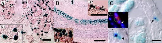
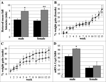
To gain insight into the expression pattern and functional importance of the forkhead transcription factor Foxs1, we constructed a Foxs1-beta-galactosidase reporter gene "knock-in" (Foxs1beta-gal/beta-gal) mouse, in which the wild-type (wt) Foxs1 allele has been inactivated and replaced by a beta-galactosidase reporter gene. Staining for beta-galactosidase activity reveals an expression pattern encompassing neural crest-derived cells, e.g., cranial and dorsal root ganglia as well as several other cell populations in the central nervous system (CNS), most prominently the internal granule layer of cerebellum. Other sites of expression include the lachrymal gland, outer nuclear layer of retina, enteric ganglion neurons, and a subset of thalamic and hypothalamic nuclei. In the CNS, blood vessel-associated smooth muscle cells and pericytes stain positive for Foxs1. Foxs1beta-gal/beta-gal mice perform significantly better (P < 0.01) on a rotating rod than do wt littermates. We have also noted a lower body weight gain (P < 0.05) in Foxs1beta-gal/lbeta-gal males on a high-fat diet, and we speculate that dorsomedial hypothalamic neurons, expressing Foxs1, could play a role in regulating body weight via regulation of sympathetic outflow. In support of this, we observed increased levels of uncoupling protein 1 mRNA in Foxs1beta-gal/beta-gal mice. This points toward a role for Foxs1 in the integration and processing of neuronal signals of importance for energy turnover and motor function.


Figures

FIG. 1.
FIG. 1.
Strategy for disruption of the Foxs1 gene. (A) Schematic representation of the Foxs1 locus, the targeting vector, and the recombinant allele. The targeting vector carried a neomycin resistance gene (neor) and an Escherichia coli lacZ gene fitted with a nuclear localization signal (nls-lacZ) that had been fused in frame with the Foxs1 coding sequence 16 amino acids downstream of its initiation codon. Homologous recombination resulted in a mutant allele where most of the Foxs1 gene, including the DNA binding region (Box), had been deleted. Arrows indicate the sizes of restriction fragments detected by the short arm (s.a.) probe in the wt and recombinant alleles. Southern blot analysis of EcoRV-digested DNA from wild-type and targeted ES cells (B) and mice (C). The short arm probe detected a 5.5-kb band for the wild-type allele and a 4-kb fragment for the targeted allele. The ES cell clone denoted 39P was used for production of mice.
FIG. 2.
FIG. 2.
Early embryonic expression pattern of Foxs1. (A) Whole-mount Foxs1 in situ hybridization using an Foxs1 cRNA probe. Transcripts were detected in the trigeminal (V), facio-acoustic (VII-VIII), superior (IXs), and jugular (Xj) ganglia and also in DRG. (B to E) Whole-mount X-Gal staining of Foxs1+/β-gal heterozygotic embryos of ages ranging from E9.5 to E12.5. Expression of Foxs1 is initiated after E8.5 and by E9.5 (B), X-Gal staining can be detected in the trigeminal ganglion and the facio-acoustic ganglia and in a few DRG. By E10.5 (C), expression has extended to DRG along the rostro-caudal axis and to the petrosal (IXp) and nodose (Xn) ganglia as well as in the nasal region of the embryo (arrows in panel C). In the E11.5 embryo (D), Foxs1 expression can be detected in the superior and jugular ganglia, and by E12.5 (E), Foxs1 expression can be detected along major cephalic blood vessels (arrow in panel E). (F) Staining in all cranial sensory ganglia evident in transverse section of E10.5 embryo. The level of the transverse section is indicated by the red line in panel C.
FIG. 3.
FIG. 3.
Late embryonic expression of Foxs1. X-Gal staining of 100-μm vibratome sections of E15.5 (A to C) and E18.5 (D to G) embryos is shown. (A) Foxs1 expression is present in the EGL of the forming cerebellum and in the deep fastigial nucleus (FN). Staining remains strong in sensory division of cranial ganglia (B) as well as in DRG, but at this stage, Foxs1 is also expressed in peripherally located Schwann cells along the ventral ramus of the spinal nerve (C). In the E18.5 embryo, cells just beneath the nasal epithelium (arrows in panel D), in the cartilage primordium of the nasal septum, stain positive for β-galactosidase. At this stage, the retina (ret) is negative for β-galactosidase (D). Expression is evident in the cochlear nucleus (CN) and in Schwann cells of the cochlear (cn) and vestibular (vn) nerves extending from the vestibulocochlear ganglion (VIII) (E) and also in Schwann cells of a branch of the trigeminal nerve (nV) extending toward follicles of vibrissae (fvib) (F). (G) Foxs1 expression has been initiated in the gray matter of the spinal cord (sc).
FIG. 4.
FIG. 4.
Sites of prominent Foxs1 expression in the P14 brain. To the left are low-magnification pictures of X-Gal-stained 200-μm coronal sections indicating the approximate level of the higher-magnification pictures, to the right, of 20-μm sections stained with both X-Gal and cresyl violet. Close-ups are depicted in black and white frames referring to sections marked in low-magnification pictures. (A) Anteroventral (AV) and anteromedial (AM) nuclei of thalamus. (B) Posterior hypothalamic area (PHA) and dorsomedial nucleus (DMH) of hypothalamus. (C) Cortical (Cx) and presubicular (PRS) layer VI neurons and periaqueductal gray matter (PAG). (D) Layer bordering the white matter in medial (MEnt) and lateral (LEnt) entorhinal cortices. (E) Fastigial (FN) and cochlear (CN) nuclei. (F) Cerebellar IGL and a subpopulation of Purkinje cells (PC, arrows) and vestibular nuclei (VN). (G) External cuneate (ECu) and lateral reticular (LRN) nuclei. Bars, 50 μm.
FIG. 5.
FIG. 5.
Cellular localization of Foxs1 expression. (A to D) Immunostaining of sagittal sections of E11.5 Foxs1β-gal/β-gal embryos with antibodies against β-galactosidase (orange), neurofilament 160 (green), and a nuclear counterstain (red). Foxs1 is expressed in most if not all of the neurons located in the trigeminal (A), vestibulocochlear (B), proximal and distal ganglia of glossopharyngeal and vagal nerves (C), and DRG (D). (E to H) Foxs1 expression in P7 dorsal root ganglia. Immunofluorescence with anti-β-galactosidase antibody (green) and a nuclear counterstain (red) reveals that Foxs1 is not restricted to neurons of a specific sensory modality but colocalizes with several different markers in P7 Foxs1β-gal/β-gal DRG. Colocalization is seen with CGRP (orange) (E), substance P (orange) (F), and parvalbumin (orange) (G). (H) β-Galactosidase-positive cells are also seen along the nerve sheet extending from the DRG. (I and J) Cerebellar expression of Foxs1 in P14 brain. (I) Low-magnification view of X-Gal-stained sagittal section identifying Foxs1 in the internal granule layer. (J) Immunofluorescence analysis with antibodies against β-galactosidase (green), calbindin D28K (red, a marker of Purkinje cells), and GABAA receptor α6 (orange, a marker of granule cells) confirms Foxs1 expression in most or all granule cells as well as in a subpopulation of Purkinje cells (arrows). Bars, 100 μm (A to D), 20 μm (E to H), 50 μm (J).
FIG. 6.
FIG. 6.
Foxs1 expression in pericytes and vSMCs of cephalic blood vessels. (A) Brain surface blood vessels of X-Gal-stained P12 mouse brain. Arteries and arterioles (arrows) appear densely stained due to extensive coverage of Foxs1-expressing vSMCs, whereas veins (arrowheads) exhibit a less-dense staining pattern. (B) Ventral view of X-Gal-stained adult brain. β-Galactosidase-positive vSMCs line the blood vessels of the entire circle of Willis surrounding the optic chiasm (oc) and major vessels extending from it, such as middle cerebral arteries (mca), posterior cerebral arteries (pca), the basilar artery (ba), and vertebral arteries (va). Also marked are the cerebellum (Cb) and pons (P). (C) Immunofluorescence with antibodies against β-galactosidase (pink), anti-smooth muscle actin (ASMA; green), and isoloectin B4 (blue), an endothelial marker. An arteriole with ASMA immunoreactivity has smaller branches that appear to be devoid of vSMCs, while pericytes stain for β-galactosidase (pink) and the endothelium stains for isoloectin B4 (blue). (D) β-Galactosidase seen in the nuclei of ASMA-positive (green) vSMCs. Bar, 20 μm.
FIG. 7.
FIG. 7.
Sites of Foxs1 expression in adult tissues. (A, A1) In adult mouse tissues, β-galactosidase staining reveals Foxs1 expression in lachrymal glands. In the retina, neurons in the external part of the outer nuclear layer (B, B1) and some ganglion neurons (B2) express Foxs1. (C) Staining can also be found in enteric ganglia (arrow). Arrowheads in inset panels C1 and C2 point to Foxs1-expressing peripherin-positive (red in panel C1) cells. Bars, 50 μm (A and C), 200 μm (B).
FIG. 8.
FIG. 8.
(A) Rotarod performance. Bars represent mean fall latencies for wt (black bars) and Foxs1β-gal/β-gal (gray bars) male (n = 7 per genotype) and female mice (n = 8 per genotype). Minutes on the rotating rod differed significantly: *, P < 0.05; **, P < 0.01 (Mann-Whitney U test). Weight curves for female (B) and male (C) wt (squares) and Foxs1β-gal/β-gal (triangles) mice. Mice were put on a high-fat diet for 13 weeks. Repeated measures of analysis of variance indicate a significantly (P < 0.05) lower percent weight gain in Foxs1β-gal/β-gal (n = 7) male mice than in wt (n = 5) male mice. No difference was seen for female mice (n = 8 per genotype). (D) Relative mRNA levels of ucp1 were measured using quantitative real-time PCR. A small but significant difference was seen between wt (n = 4) (black bars) and Foxs1β-gal/β-gal (n = 3) (gray bars) male mice, whereas there was no difference in female mice (n = 4 per genotype). *, P < 0.05 (Student's t test). Error bars (all panels) indicate standard errors of the means.

Similar articles

Cited by

References

    1. Accili, D., and K. C. Arden. 2004. FoxOs at the crossroads of cellular metabolism, differentiation, and transformation. Cell 117:421-426. - PubMed
    1. Ang, S. L., and J. Rossant. 1994. HNF-3 beta is essential for node and notochord formation in mouse development. Cell 78:561-574. - PubMed
    1. Bergquist, F., H. N. Shahabi, and H. Nissbrandt. 2003. Somatodendritic dopamine release in rat substantia nigra influences motor performance on the accelerating rod. Brain Res. 973:81-91. - PubMed
    1. Bernardis, L. L. 1975. The dorsomedial hypothalamic nucleus in autonomic and neuroendocrine homeostasis. Can. J. Neurol. Sci. 2:45-60. - PubMed
    1. Bernardis, L. L., and L. L. Bellinger. 1991. Brown (BAT) and white (WAT) adipose tissue in high-fat junk food (HFJF) and chow-fed rats with dorsomedial hypothalamic lesions (DMNL rats). Behav. Brain Res. 43:191-195. - PubMed

Publication types

MeSH terms