Comparative Study
. 2005 Jul;25(13):5763-76.
doi: 10.1128/MCB.25.13.5763-5776.2005.

Generation and characterization of Rac3 knockout mice

Affiliations
Comparative Study

Generation and characterization of Rac3 knockout mice

Sara Corbetta et al. Mol Cell Biol. 2005 Jul.

Abstract

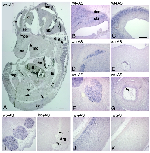
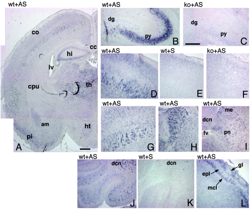
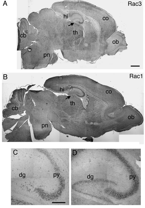
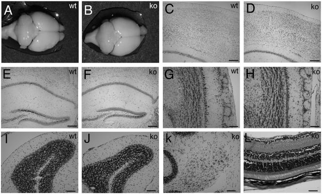
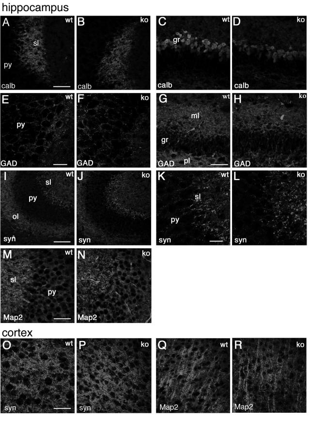
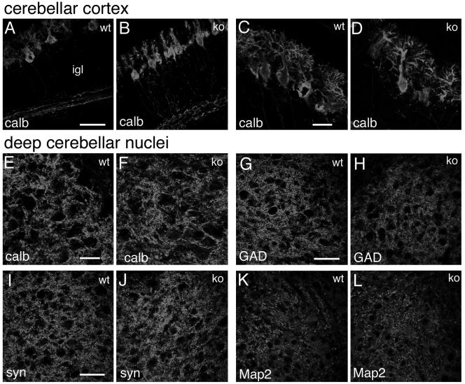
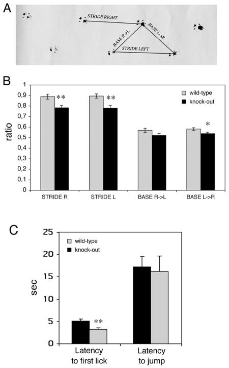
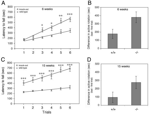
Rac proteins are members of the Rho family of GTPases involved in the regulation of actin dynamics. The three highly homologous Rac proteins in mammals are the ubiquitous Rac1, the hematopoiesis-specific Rac2, and the least-characterized Rac3. We show here that Rac3 mRNA is widely and specifically expressed in the developing nervous system, with highest concentration at embryonic day 13 in the dorsal root ganglia and ventral spinal cord. At postnatal day 7 Rac3 appears particularly abundant in populations of projection neurons in several regions of the brain, including the fifth layer of the cortex and the CA1-CA3 region of the hippocampus. We generated mice deleted for the Rac3 gene with the aim of analyzing the function of this GTPase in vivo. Rac3 knockout animals survive embryogenesis and show no obvious developmental defects. Interestingly, specific behavioral differences were detected in the Rac3-deficient animals, since motor coordination and motor learning on the rotarod was superior to that of their wild-type littermates. No obvious histological or immunohistological differences were observed at major sites of Rac3 expression. Our results indicate that, in vivo, Rac3 activity is not strictly required for normal development in utero but may be relevant to later events in the development of a functional nervous system.


Figures

FIG. 1.
FIG. 1.
Targeted disruption of the mouse Rac3 gene. (A) Targeting strategy. The structures of the wild-type allele and the disrupted allele are shown. In the wild-type allele, exons are numbered 1 to 6 and are represented by black boxes. The primers used for routine genotyping are represented by arrows. (B) PCR genotyping with primers for wild-type and recombinant alleles. A 0.46-kb fragment was generated from the wild-type allele (primers F1 and R2), and a 0.37 kb fragment was generated from the mutant allele (primers F1 and R3). (C) Genomic DNA prepared from tail biopsies was used for Southern blot analysis after digestion with EcoRI and XbaI (upper blot), or with SacI (lower blot). Filters were probed with the 0.25-kb EcoRI/SacI fragment generated from sequence immediately upstream of the 5′ arm (5′ probe, upper blot), and with the 0.4-kb KpnI/SmaI fragment from the 3′ arm (3′ probe, lower blot). ATG, start methionine; lp, loxP sites; +/+, wild-type; +/−, heterozygous; −/−, homozygous knockout.
FIG. 2.
FIG. 2.
Analysis of the expression of the transcripts of Rac proteins in mutant mice. (A) Total RNA prepared from P9 brains of wild-type (+/+), heterozygous (+/−), and knockout (−/−) mice was reverse transcribed and then used for PCR amplification with primers specific for Rac1 and Rac3. No amplification product for Rac3 was observed in the knockout mice. Total RNA (15 μg/lane) from P0 (B and D) or P9 (C and E) brains of wild-type (+/+), heterozygous (+/−), and knockout (−/−) mice was used for Northern blot analysis. Filters were incubated at high stringency with a 0.28-kb Rac3-specific probe (B), a 0.38-kb Rac2-specific probe (C), the 1-kb Rac3-specific probe also used for in situ hybridization (D), or a 1.2-kb Rac1-specific probe (E). A control lane with 15 μg of total RNA from adult spleen was included in panel C. No RNA for Rac3 was detectable in the knockout mice (B and D).
FIG. 3.
FIG. 3.
Expression of the Rac3 protein. (A) Immunoblotting on P9 brain lysates. Filters were incubated with the anti-Rac3 (upper), anti-Rac1 (middle), or antitubulin (lower) antibodies. No Rac3 protein was detected in the brain lysate from knockout mice. (B) P9 brain lysates were incubated with protein A-Sepharose beads conjugated to preimmune (PI) or immune anti-Rac3 serum (IP Rac3). After blotting, the filters were incubated with the anti-Rac monoclonal antibody. No Rac3 protein was immunoprecipitated from the brain lysate of knockout mice. (C) Aliquots of 2 mg protein of P7 brain lysate and of 3 mg protein of adult tissue lysates were immunoprecipitated with anti-Rac3 antibody. After blotting, the filters were incubated with the anti-Rac monoclonal antibody recognizing both Rac3 and Rac1. Br, brain; Sp, spleen; Li, liver; Lu, lung; Mu, skeletal muscle; Ki, kidney; Cb, cerebellum; He, heart; Te, testis; Pa, pancreas; Th, thymus. (D) Aliquots of lysates from human platelets (1.12 mg protein), P7 brain (2 mg protein), and mouse peritoneal macrophages (1.2 mg protein) were immunoprecipitated with anti-Rac3 (IP) or with preimmune serum (PI) and blotted with the anti-Rac monoclonal antibody. No Rac3 could be detected in platelets and macrophages. Ub, unbound material after immunoprecipitation (250 μg/lane); Ly, lysate (100 μg/lane).
FIG. 4.
FIG. 4.
Expression of Rac3 mRNA in E13 mice. Parasagittal sections of E12.5 to E13 wild-type (wt) and knockout (ko) mouse embryos were hybridized with antisense (AS) or sense (S) digoxigenin-labeled cRNAs for Rac3 and examined for signal detection. (A) Rac3 is specifically and widely expressed in the developing nervous system. (B) Cerebellum; (C) mesencephalon; (D and E) medulla oblongata; (F) trigeminal ganglion; (G) eye; (H and I) dorsal root ganglia; (J and K) spinal cord. Abbreviations: ctz, cortical transitory zone; dcn, deep cerebellar nuclei; di, diencephalon; drg, dorsal root ganglia; hb, hindbrain; he, heart; li, liver; lu, lung; mc, mouth cavity; me, mesencephalon; mg, midgut; nc, nasal cavity; ob, olfactory bulb; re, rectum; sc, spinal cord; se, septum; te, telencephalic vesicle. Bars: 500 μm (A); 200 μm (B to K).
FIG. 5.
FIG. 5.
Expression of Rac3 mRNA in P7 mouse brain. In situ hybridization on P7 mouse brain coronal (A, C, F to H) and parasagittal (B, D, E, I to L) sections from wild-type (wt) and knockout (ko) mice, using a Rac3 antisense (AS) or sense (S) probe. (A) Rac3 is specifically expressed in several areas of the P7 brain. (B and C) hippocampus; (D to G) cerebral cortex; (H) thalamus; (I) pons; (J and K) cerebellum; (L) olfactory bulb. Abbreviations: am, amygdala; cc, corpus callosum; co, cortex; cpu, caudatus-putamen; dcn, deep cerebellar nuclei; dg, dentate gyrus; epl, external plexiform layer; fv, fourth ventricle; gl, glomerular layer of olfactory bulb; hi, hippocampus; ht, hypothalamus; lv, lateral ventricle; mcl, mitral cell layer; me, mesencephalon; pi, piriform cortex; pn, pons; py, pyramidal cell layer of the hippocampus; th, thalamus. Bars: 500 μm (A); 200 μm (B to L).
FIG. 6.
FIG. 6.
Expression of Rac1 and Rac3 mRNA in P7 mouse brain. In situ hybridization on P7 mouse brain parasagittal sections from wild-type mice, using Rac3 (A and C) or Rac1 (B and D) antisense probes, as detailed in the text. Abbreviations: cb, cerebellum; co, cortex; dg, dentate gyrus; hi, hippocampus; ob, olfactory bulb; pn, pons; py, pyramidal cell layer of the hippocampus; th, thalamus. Arrows in A and B indicate an evident difference in labeling in the dentate gyrus between Rac3 and Rac1. Bars: 500 μm (A and B); 200 μm (C and D).
FIG. 7.
FIG. 7.
Histology of wild-type and knockout adult brains. Macroscopic analysis of brains from 3-month-old wild-type (A) and knockout (B) mice revealed no major abnormalities. Coronal sections stained with cresyl violet of brains through cerebral cortex (C and D), hippocampus (E and F), olfactory bulbs (G and H), cerebellar cortex (I and J), and deep cerebellar nuclei (K) showed normal cell layering and an unaltered cytoarchitecture in both wild-type and knockout mice. (L) Section through knockout adult retina stained with hematoxylin and eosin, showing normal layering. Bars: 200 μm (C to F), 100 μm (G to K), 50 μm (L).
FIG. 8.
FIG. 8.
Distribution of neuronal markers in the hippocampus and in the cortex of P7 mice. Immunofluorescence on P7 brain coronal sections from wild-type (wt) and knockout (ko) mice, incubated with the indicated primary antibodies, and Alexa-488-labeled secondary antibodies. (A to N) Hippocampus; (O to R) fourth to sixth layers of the cerebral cortex. Abbreviations: gr, granular layer of dentate gyrus; ml, molecular layer of dentate gyrus; ol, oriens layer of the CA3 region; sl, stratum lucidem; pl, polymorph layer of dentate gyrus; py, pyramidal cell layer of the CA3 region; calb, calbindin; syn, synapsin I; GAD, GAD65 and GAD67 staining. Bars: 100 μm (I and J); 50 μm (A to D, G, H, M, and N); 25 μm (E, F, K, and L).
FIG. 9.
FIG. 9.
Distribution of neuronal markers in the cerebellum of P7 mice. Immunofluorescence on P7 cerebellar parasagittal sections from wild-type (wt) and knockout (ko) mice, incubated with the indicated primary antibodies and Alexa-488-labeled secondary antibodies. (A to D) Cerebellar cortex; (E to L) deep cerebellar nuclei. Abbreviations: P, Purkinje cells; igl, inner granular layer. Bars: 50 μm (A, B, and G to L); 25 μm (C to F).
FIG. 10.
FIG. 10.
Behavioral characterization of wild-type and Rac3 knockout mice: analysis of footprint patterns and hot plate test. (A) Experimental set-up used for footprint analysis. Stride right, stride left, base R>L (right to left) and base L>R (left to right) indicate the four parameters considered. (B) Footprint patterns of 24- to 28-week-old wild-type (n = 9, grey bars) and knockout (n = 9, black bars) mice were quantitatively assessed for stride R, stride L, base R>L, and base L>R, as detailed in Materials and Methods. The mean values (± standard error of the mean) normalized by the body length are shown. Rac3 knockout animals had slightly decreased strides. (C) Ten wild-type and 10 knockout mice were placed on a hot plate at 55°C. Latency to first paw lick and latency to jump were recorded. Each bar shows the mean ± standard error of the mean. *, P < 0.05; **, P < 0.005.
FIG. 11.
FIG. 11.
Rotarod learning is improved in Rac3 knockout mice. (A) Time that mice remained on the rotarod before falling as a function of training session of 6-week-old animals. Open circles represent wild-type Rac3+/+ (n = 10) and open squares represent Rac3−/− mice (n = 10). (C) Performance on the accelerating Rotarod was tested on the same groups of mice at 15 weeks. Each point represents the mean of three tests. Lines of best fit had a correlation coefficient of 0.92 or greater. Error bars indicate standard error of the mean. Statistical significance in panels A and C: *, P < 0.005; **, P < 0.001; ***, P < 0,0005. (B) Motor learning for animals at 6 weeks and (D) 15 weeks of age. Bars represent differences in active rotation behavior (trial 6 − trial 1) for wild-type (+/+) and knockout (−/−) animals (P < 0.05 in panels B and D).

References

    1. Abdel-Latif, D., M. Steward, D. L. Macdonald, G. A. Francis, M. C. Dinauer, and P. Lacy. 2004. Rac2 is critical for neutrophil primary granule exocytosis. Blood 104:832-839. - PubMed
    1. Abo, A., E. Pick, A. Hall, N. Totty, C. G. Teahan, and A. W. Segal. 1991. Activation of the NADPH oxidase involves the small GTP-binding protein p21rac1. Nature 353:668-670. - PubMed
    1. Albertinazzi, C., D. Gilardelli, S. Paris, R. Longhi, and I. de Curtis. 1998. Overexpression of a neural-specific Rho family GTPase, cRac1B, selectively induces enhanced neuritogenesis and neurite branching in primary neurons. J. Cell Biol. 142:815-825. - PMC - PubMed
    1. Ando, S., K. Kaibuchi, T. Sasaki, K. Hiraoka, T. Nishiyama, T. Mizuno, M. Asada, H. Nunoi, I. Matsuda, Y. Matsuura, P. Polakis, F. McCormick, and Y. Takai. 1992. Post-translational processing of Rac p21s is important both for their interaction with the GDP/GTP exchange proteins and for their activation of NADPH oxidase. J. Biol. Chem. 267:25709-25713. - PubMed
    1. Benvenuti, F., S. Hugues, M. Walmsley, S. Ruf, L. Fetler, M. Popoff, V. L. Tybulewicz, and S. Amigorena. 2004. Requirement of Rac1 and Rac2 expression by mature dendritic cells for T cell priming. Science 305:1150-1153. - PubMed

Publication types

MeSH terms