. 2005 Jul;25(13):5777-85.
doi: 10.1128/MCB.25.13.5777-5785.2005.

Generation of rac3 null mutant mice: role of Rac3 in Bcr/Abl-caused lymphoblastic leukemia

Affiliations

Generation of rac3 null mutant mice: role of Rac3 in Bcr/Abl-caused lymphoblastic leukemia

Young Jin Cho et al. Mol Cell Biol. 2005 Jul.

Abstract

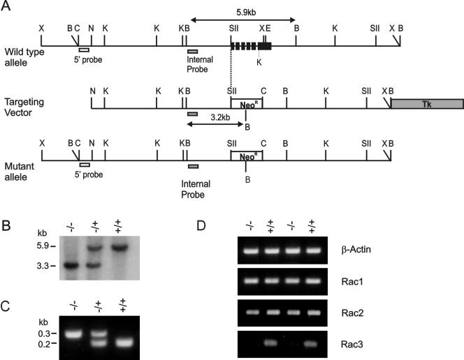
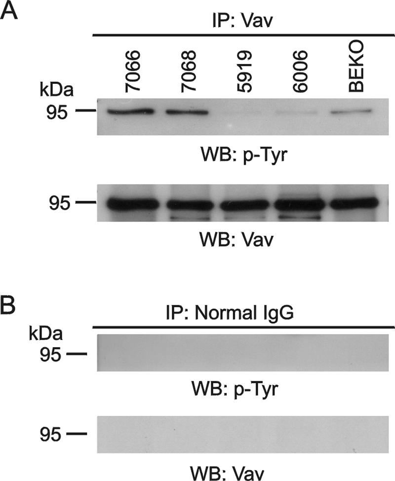
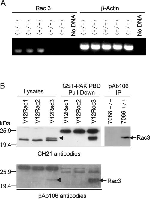
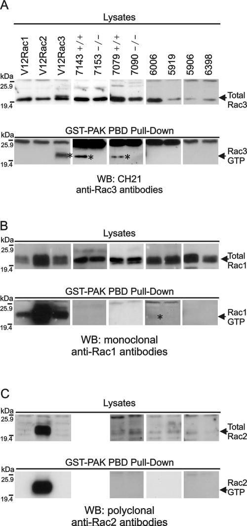
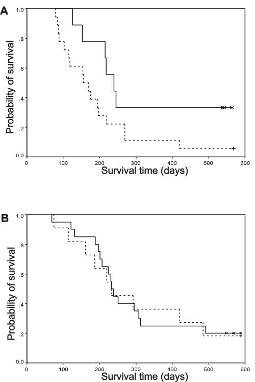
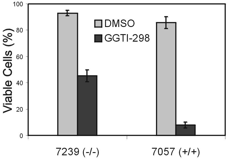
Numerous studies indirectly implicate Rac GTPases in cancer. To investigate if Rac3 contributes to normal or malignant cell function, we generated rac3 null mutants through gene targeting. These mice were viable, fertile, and lacked an obvious external phenotype. This shows Rac3 function is dispensable for embryonic development. Bcr/Abl is a deregulated tyrosine kinase that causes chronic myelogenous leukemia and Ph-positive acute lymphoblastic leukemia in humans. Vav1, a hematopoiesis-specific exchange factor for Rac, was constitutively tyrosine phosphorylated in primary lymphomas from Bcr/Abl P190 transgenic mice, suggesting inappropriate Rac activation. rac3 is expressed in these malignant hematopoietic cells. Using lysates from BCR/ABL transgenic mice that express or lack rac3, we detected the presence of activated Rac3 but not Rac1 or Rac2 in the malignant precursor B-lineage lymphoblasts. In addition, in female P190 BCR/ABL transgenic mice, lack of rac3 was associated with a longer average survival. These data are the first to directly show a stimulatory role for Rac in leukemia in vivo. Moreover, our data suggest that interference with Rac3 activity, for example, by using geranyl-geranyltransferase inhibitors, may provide a positive clinical benefit for patients with Ph-positive acute lymphoblastic leukemia.


Figures

FIG. 1.
FIG. 1.
Generation of mice deficient in rac3. (A) Schematic illustration of the wild-type rac3 genomic locus (top line), the targeting vector (middle line), and the correctly targeted allele (bottom line). The targeting vector was designed to replace a 1.8-kb SstII-Eco47III fragment with a PGK-neomycin resistance cassette. The grey box in the targeting vector indicates the location of the herpes simplex virus thymidine kinase gene and plasmid sequences. The open box shows the location of the PGK-neo cassette. We used an 0.4-kb ClaI-NruI fragment from outside the targeting vector as an external probe, indicated as 5′ probe beneath the maps. The approximate locations of the six exons of mouse rac3 are indicated in the wild-type allele as black boxes. Restriction enzymes: B = BamHI; C = ClaI; E = Eco47III; K = KpnI; N = Nru I; SII = SstII; Xb = XbaI. (B and C) Genotyping was done using Southern blot analysis on BamHI-digested genomic DNA from the F9 progeny hybridized to the internal probe indicated in panel A (B) or using PCR (C). (D) RT-PCR on two different sets of rac3−/− and rac3+/+ bone marrow-derived macrophages shows that no rac3 mRNA is made in rac3−/− samples. The primers used are indicated in Table 1.
FIG. 2.
FIG. 2.
Vav is tyrosine phosphorylated in primary P190 lymphoblastic leukemia cells. (A) Vav was immunoprecipitated (IP) with anti-Vav antibodies from lymphoma lysates of mice 7066 (P190 BCR/ABL rac3+/+); 7068 (P190 BCR/ABL rac3−/−); 5919 (wild type), 6006 (10xCRKL transgenic); and BEKO (bcr−/−). The samples were separated on a 7.5% SDS-polyacrylamide gel, and the presence of phosphorylated Vav (top) and total Vav (bottom) was investigated with antiphosphotyrosine antibodies (PY-20) or anti-Vav antibodies (C-14). (B) Control immunoprecipitation with immunoglobulin G. The Western blot (WB) was reacted with the antibodies shown.
FIG. 3.
FIG. 3.
Detection of Rac3 expression. (A) RT-PCR of rac3 mRNA in leukemic precursor B cells. The primers used are indicated above the lanes. rac3+/+ and rac3−/− indicate samples from P190 rac3+/+ and P190 rac3−/− mice, respectively. (B) Western blot analysis using two different Rac3 antibodies (as indicated beneath the panels) to detect Rac3. CHO cells were transfected with constitutively active (V12) Rac1, Rac2, or Rac3, and activated Rac was detected using GST-Pak. Total Rac3 protein was also immunoprecipitated from lymphoma lysates of a rac3−/− and a rac3+/+ mouse using pAb106 antibodies. The location of Rac3 protein is indicated.
FIG. 4.
FIG. 4.
Analysis of Rac activation in primary lymphomas, for activated Rac3 using anti-CH21 antibodies (A); for activated Rac1 using monoclonal anti-Rac1 (Transduction Laboratories) antibodies (B); and for activated Rac2 using anti-Rac2 (Santa Cruz) antibodies (C). Top panels, lysates for total Rac levels; bottom panels, GST-Pak-RBD pull-down reactions for GTP-bound Rac. COS cells transfected with V12Rac1, Rac2, or Rac3 are shown in the three left lanes as positive methodological controls. All samples were lymphomas (unless otherwise indicated) and include 7143 (P190 BCR/ABL rac3+/+); 7153 (P190 BCR/ABL rac3−/−); 7079 (P190 BCR/ABL rac3+/+); 7090 (P190 BCR/ABL rac3−/−); 6006 (10xCRKL transgenic); 5919 (wild type); 5906 (wild type, benign mammary gland tumor); and 6398 (10xCRKL transgenic). Different exposures of the same blot are shown for lysates and GST-Pak-RBD pull-downs. The position of activated GTP-bound Rac (RacGTP) is indicated with an asterisk.
FIG. 5.
FIG. 5.
Kaplan-Meier survival curve of P190 BCR/ABL transgenic mice in the presence and absence of rac3. (A) A genetically matched set of P190 BCR/ABL rac3+/+ female mice (dashed line, n = 18) were compared to P190 BCR/ABL rac3−/− female mice (solid line, n = 9). (B) A matched set of P190 BCR/ABL rac3+/+ male mice (dashed line, n = 20) were compared to P190 BCR/ABL rac3−/− male mice (solid line, n = 11). Censored data are marked (asterisks and cross).
FIG. 6.
FIG. 6.
P190 BCR/ABL precursor B lymphoblasts expressing Rac3 are more sensitive to GGTI treatment than cells lacking Rac3. Lymphoblasts grown on mitotically inactivated murine embryonic fibroblast feeder layers were treated for 24 h with 20 μM GGTI-298 in dimethyl sulfoxide or dimethyl sulfoxide alone, after which the percentage of viable cells was determined. The result shown is one of two independently performed experiments with similar results. A second set of rac3−/− and rac3+/+ lymphoblasts yielded a qualitatively similar result. Bars indicate the standard error of the mean.

References

    1. Aghazadeh, B., W. E. Lowry, X. Y. Huang, and M. K. Rosen. 2000. Structural basis for relief of autoinhibition of the Dbl homology domain of proto-oncogene Vav by tyrosine phosphorylation. Cell 102:625-633. - PubMed
    1. Bassermann, F., T. Jahn, C. Miething, P. Seipel, R. Y. Bai, S. Coutinho, V. L. Tybulewicz, C. Peschel, and J. Duyster. 2002. Association of Bcr-Abl with the proto-oncogene Vav is implicated in activation of the Rac-1 pathway. J. Biol. Chem. 277:12437-12445. - PubMed
    1. Benard, V., B. P. Bohl, and G. M. Bokoch. 1999. Characterization of Rac and Cdc42 activation in chemoattractant-stimulated human neutrophils using a novel assay for active GTPases. J. Biol. Chem. 274:13198-13204. - PubMed
    1. Bolis, A., S. Corbetta, A. Cioce, and I. de Curtis. 2003. Differential distribution of Rac1 and Rac3 GTPases in the developing mouse brain: implications for a role of Rac3 in Purkinje cell differentiation. Eur. J. Neurosci. 18:2417-2424. - PubMed
    1. Crespo, P., K. E. Schuebel, A. A. Ostrom, J. S. Gutkind, and X. R. Bustelo. 1997. Phosphotyrosine-dependent activation of Rac-1 GDP/GTP exchange by the Vav proto-oncogene product. Nature 385:169-172. - PubMed

Publication types

MeSH terms

Substances