. 2005 Jun 15;19(12):1432-7.
doi: 10.1101/gad.1299505.

Bmi-1 promotes neural stem cell self-renewal and neural development but not mouse growth and survival by repressing the p16Ink4a and p19Arf senescence pathways

Affiliations

Bmi-1 promotes neural stem cell self-renewal and neural development but not mouse growth and survival by repressing the p16Ink4a and p19Arf senescence pathways

Anna V Molofsky et al. Genes Dev. .

Abstract

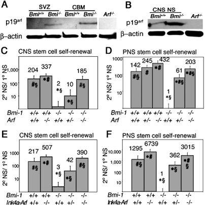
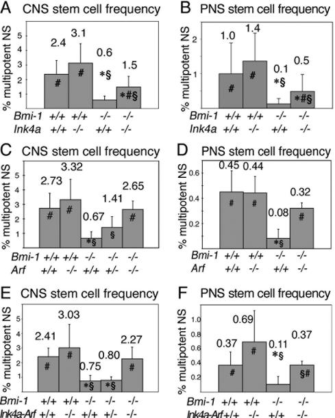
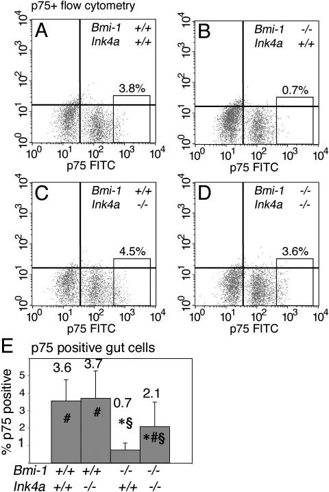
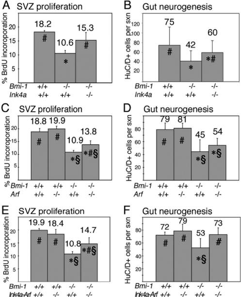
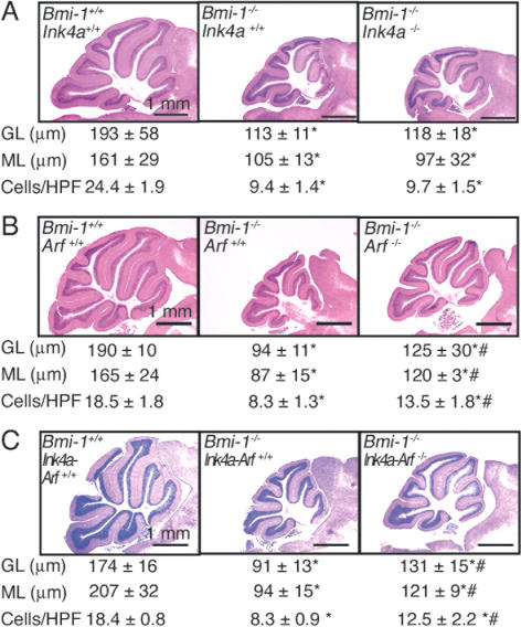
Bmi-1 is required for the post-natal maintenance of stem cells in multiple tissues including the central nervous system (CNS) and peripheral nervous system (PNS). Deletion of Ink4a or Arf from Bmi-1(-/-) mice partially rescued stem cell self-renewal and stem cell frequency in the CNS and PNS, as well as forebrain proliferation and gut neurogenesis. Arf deficiency, but not Ink4a deficiency, partially rescued cerebellum development, demonstrating regional differences in the sensitivity of progenitors to p16Ink4a and p19Arf. Deletion of both Ink4a and Arf did not affect the growth or survival of Bmi-1(-/-) mice or completely rescue neural development. Bmi-1 thus prevents the premature senescence of neural stem cells by repressing Ink4a and Arf, but additional pathways must also function downstream of Bmi-1.


Figures

Figure 1.
Figure 1.
p19Arf is up-regulated in the absence of Bmi-1, and deletion of Arf or Ink4a-Arf significantly increased the self-renewal of neural stem cells from the SVZ and gut of Bmi-1-/- mice. (A) p19Arf was increased in uncultured SVZ and cerebellum (CBM) of 4-wk-old Bmi-1-/- mice. Western blot of neurosphere cells from Arf-/- mice is shown as a negative control. (B) p19Arf was increased in Bmi-1-/- CNS neurospheres (NS) cultured for 13 d. (C,D) Arf deficiency significantly increased self-renewal (the number of secondary neurospheres generated per subcloned primary neurosphere) within Bmi-1+/+ and Bmi-1-/- CNS neurospheres (C) and PNS neurospheres (D), as well as the percentage of cells from Bmi-1-/- (but not Bmi-1+/+) primary neurospheres that formed secondary neurospheres upon replating (Supplementary Fig. 6). CNS data represent mean ± SD for two to four mice per genotype, in four independent experiments. PNS data are from three to seven mice per genotype in four independent experiments. (E,F) Ink4a-Arf deficiency significantly increased self-renewal within Bmi-1+/+ and Bmi-1-/- CNS neurospheres (E; mean ± SD for two to four mice per genotype, four independent experiments) and PNS neurospheres (F; three to six mice per genotype, six independent experiments), and the percentage of cells from Bmi-1-/- (but not Bmi-1+/+) primary neurospheres that formed secondary neurospheres upon replating (Supplementary Fig. 6). () Significantly different (P < 0.05) from Bmi-1+/+Arf+/+ or Bmi-1+/+Ink4a-Arf+/+; (#) significantly different from Bmi-1-/-Arf+/+ or Bmi-1-/-Ink4a-Arf+/+; (§) significantly different from Bmi-1+/+Arf-/- or Bmi-1+/+Ink4a-Arf-/-. All experiments were performed on 4- to 8-wk-old mice.
Figure 2.
Figure 2.
Deletion of Ink4a, Arf, or Ink4a-Arf significantly increased neural stem cell frequency in Bmi-1-/- mice. (A,B) Ink4a deficiency significantly increased the percentage of SVZ cells (A) or adult gut NCSCs (B) from Bmi-1-/- mice that formed multipotent neurospheres in culture (mean ± SD for four independent experiments). (C,D) Arf deficiency also significantly increased the percentage of Bmi-1-/- cells that formed multipotent neurospheres from the SVZ (C; three to six mice per genotype, in six independent experiments) or gut wall (D; four mice per genotype, in four independent experiments). (E,F) Ink4a-Arf deficiency significantly increased the frequency of cells from the Bmi-1-/- adult SVZ (E; two to seven mice per genotype, eight independent experiments) or gut wall (F; three to eight mice per genotype, three independent experiments) that formed multipotent neurospheres in culture. () Significantly different (P < 0.05 by t-test) from wild-type; (#) significantly different from Bmi-1-/-Ink4a+/+, Bmi-1-/-Arf+/+, or Bmi-1-/-Ink4a-Arf+/+; (§) significantly different from Bmi-1+/+Ink4a-/-, Bmi-1+/+Arf-/-, or Bmi-1+/+Ink4a-Arf-/-. All experiments employed 4- to 8-wk-old mice.
Figure 3.
Figure 3.
Ink4a deficiency partially rescued the frequency of uncultured NCSCs in the adult gut. Ink4a deficiency partially rescued the frequency of p75+ cells, which are highly enriched for NCSCs, in the adult gut wall of Bmi-1-/- mice. (A-D) Representative flow-cytometry plots from the indicated genotypes demonstrating p75+ NCSC frequency (boxed region of each plot) among freshly dissociated gut wall cells. (E) The frequency of p75+ cells was significantly reduced by Bmi-1 deficiency, and significantly increased by Ink4a deficiency. Data in panel E represent mean ± SD for 10 independent experiments using 4- to 8-wk-old mice. Statistical significance is indicated as in Figure 2.
Figure 4.
Figure 4.
Deletion of Ink4a, Arf, or Ink4a-Arf partially rescued SVZ proliferation and gut neurogenesis in 3- to 9-wk-old Bmi-1-/- mice in vivo. (A,C,E) The rate of proliferation (percentage of cells that incorporate a pulse of BrdU) in the SVZ of Bmi-1-/- mice was significantly increased by Ink4a deficiency (A), Arf deficiency (C), or combined Ink4a-Arf deficiency (E). Note that deletion of these genes had no effect on SVZ proliferation in Bmi-1+/+ mice (C,E; Ink4a data not shown; six to eight sections per mouse, three to five mice per genotype). Ink4a deficiency partially rescued the number of myenteric plexus neurons per cross-section through the distal small intestine of Bmi-1-/- mice (B; mean ± SD for four to seven mice per genotype, seven to 10 sections per mouse). Arf deficiency tended to increase the number of neurons per cross-section in Bmi-1-/- mice, though the effect was not statistically significant (D; mean ± SD for four to five mice per genotype and eight to 10 sections per mouse). Ink4a-Arf deficiency significantly increased the number of myenteric plexus neurons per cross-section of Bmi-1-/- mice in a way that was consistent with a complete rescue (F; mean ± SD for five mice per genotype and six to eight sections per mouse). () Significantly different (P < 0.05 by t-test) from wild-type; (#) significantly different from Bmi-1-/-Ink4a+/+, Bmi-1-/-Arf+/+, or Bmi-1-/-Ink4a-Arf+/+; (§) significantly different from Bmi-1+/+Arf-/- or Bmi-1+/+Ink4a-Arf-/-.
Figure 5.
Figure 5.
Deletion of Arf or Ink4a-Arf, but not Ink4a alone, partially rescues cerebellum development in Bmi-1-/- mice. Hematoxylin and eosin-stained saggital sections of adult cerebellum. Measurements of granular layer thickness (GL), molecular layer thickness (ML), and cell density in the molecular layer (cells/high power field [HPF] is 100 μm2) for each genotype are represented below the corresponding picture. (A) Ink4a deficiency did not affect cerebellum development in Bmi-1-/- mice (mean ± SD for four mice per genotype, seven to 25 measurements per mouse). (B) Arf deficiency partially rescued cerebellum growth in 4- to 8-wk-old Bmi-1-/- mice (mean ± SD for three to four mice per genotype and 10-23 measurements per mouse). (C) Ink4a-Arf deficiency also partially rescued cerebellum growth in Bmi-1-/- mice (mean ± SD for three to five mice per genotype, six to 32 measurements per mouse). Ink4a, Arf, and Ink4a-Arf deficiencies did not affect cerebellum growth in Bmi-1+/+ mice (data not shown). Statistical significance is indicated as in Figure 4.

References

    1. Bixby S., Kruger, G.M., Mosher, J.T., Joseph, N.M., and Morrison, S.J. 2002. Cell-intrinsic differences between stem cells from different regions of the peripheral nervous system regulate the generation of neural diversity. Neuron 35: 643-656. - PubMed
    1. Bruggeman S.W.M., Valk-Lingbeek, M.E., van der Stoop, P.P.M., Jacobs, J.J.L., Kieboom, K., Tanger, E., Hulsman, D., Leung, C., Arsenijevic, Y., Marino, S., and van Lohuizen, M. 2005. Ink4a and Arf differentially affect cell proliferation and neural stem cell self-renewal in Bmi1-deficient mice. Genes & Dev. (this issue). - PMC - PubMed
    1. Dimri G.P., Martinez, J.L., Jacobs, J.J., Keblusek, P., Itahana, K., van Lohuizen, M., Campisi, J., Wazer, D.E., and Band, V. 2002. The Bmi-1 oncogene induces telomerase activity and immortalizes human mammary epithelial cells. Cancer Res. 62: 4736-4745. - PubMed
    1. Harrison D.E. 1979. Proliferative capacity of erythropoietic stem cell lines and aging: An overview. Mech. Ageing Dev. 9: 409-426. - PubMed
    1. Itahana K., Zou, Y., Itahana, Y., Martinez, J.-L., Beausejour, C., Jacobs, J.J.L., van Lohuizen, M., Band, V., Campisi, J., and Dimri, G.P. 2003. Control of the replicative life span of human fibroblasts by p16 and the polycomb protein Bmi-1. Mol. Cell. Biol. 23: 389-401. - PMC - PubMed

Publication types

MeSH terms

LinkOut - more resources