

Abnormal crossing of the optic fibres shown by evoked magnetic fields in patients with ocular albinism with a novel mutation in the OA1 gene
- PMID: 15965158
- PMCID: PMC1772728
- DOI: 10.1136/bjo.2004.060582
Abnormal crossing of the optic fibres shown by evoked magnetic fields in patients with ocular albinism with a novel mutation in the OA1 gene
Abstract
Aim: To perform genealogical and clinical studies in Finnish families with X linked ocular albinism (OA1), including characterisation of the potential misrouting of optic fibres by evaluating visual evoked magnetic fields (VEFs), and to determine the mutation behind the disease.
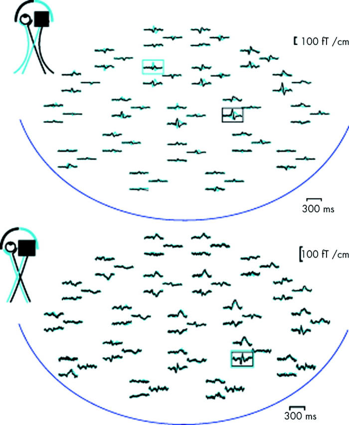
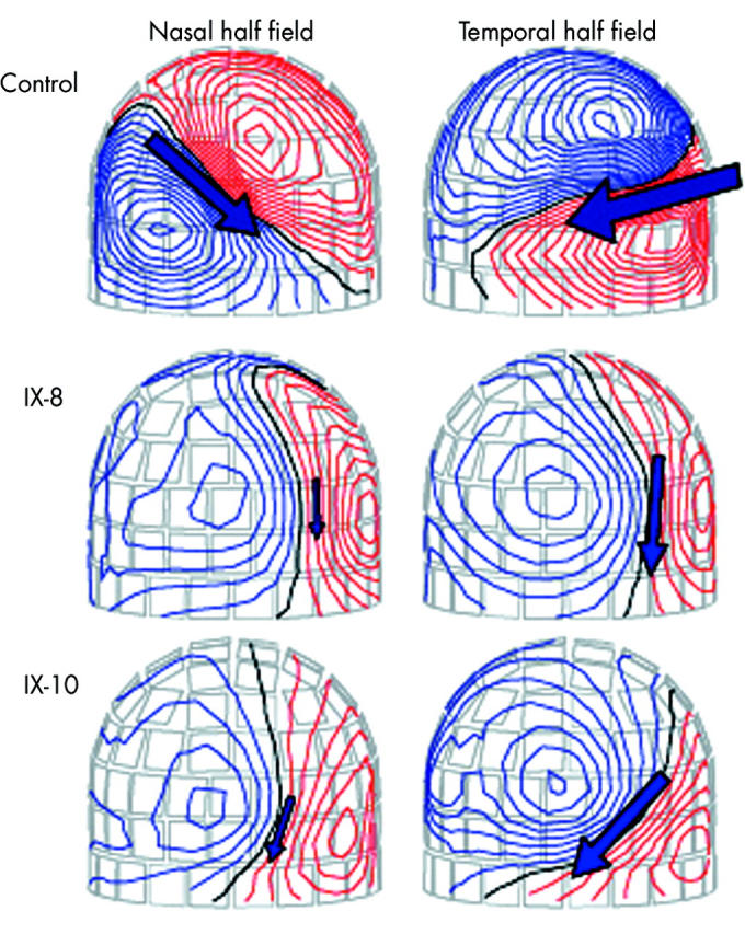
Methods: Three families with OA1 were clinically examined. VEFs were measured in two affected males and in one female carrier to characterise the cortical activation pattern after monocular visual stimulation. The neuronal sources of the VEFs were modelled with equivalent current dipoles (ECDs) in a spherical head model. All coding exons of the OA1 gene were screened for mutations by single strand conformation analysis and direct polymerase chain reaction sequencing.
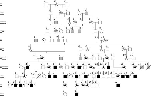
Results: Genealogical studies revealed that the three families were all related. The affected males had foveal hypoplasia with reduced visual acuity varying from 20/200 to 20/50, variable nystagmus, iris transillumination, and hypopigmentation of the retinal pigment epithelium. The ECD locations corresponding to the VEFs revealed abnormal crossing of the optic fibres in both affected males, but not in the carrier female. A novel point mutation, leading to a STOP codon, was identified in the fifth exon of the OA1 gene.
Conclusions: The data indicate that the novel mutation 640C>T in the OA1 gene is the primary cause of the eye disease in the family studied. VEFs with ECD analysis was successfully used to demonstrate abnormal crossing of the optic fibres.
Figures
Similar articles
-
New insights into ocular albinism type 1 (OA1): Mutations and polymorphisms of the OA1 gene.Hum Mutat. 2002 Feb;19(2):85-92. doi: 10.1002/humu.10034. Hum Mutat. 2002. PMID: 11793467 Review.
-
Identification of two novel mutations in families with X-linked ocular albinism.Mol Vis. 2007 Oct 2;13:1856-61. Mol Vis. 2007. PMID: 17960122
-
GPR143 mutational analysis in two Italian families with X-linked ocular albinism.Genet Test Mol Biomarkers. 2009 Aug;13(4):527-31. doi: 10.1089/gtmb.2009.0030. Genet Test Mol Biomarkers. 2009. PMID: 19604113
-
Scanning the ocular albinism 1 (OA1) gene for polymorphisms in congenital nystagmus by DHPLC.Ophthalmic Genet. 2006 Jun;27(2):43-9. doi: 10.1080/13816810600677834. Ophthalmic Genet. 2006. PMID: 16754205
-
Ocular albinism type 1: more than meets the eye.Pigment Cell Res. 2001 Aug;14(4):243-8. doi: 10.1034/j.1600-0749.2001.140403.x. Pigment Cell Res. 2001. PMID: 11549106 Review.
Cited by
-
Identification of a novel GPR143 mutation in a large Chinese family with congenital nystagmus as the most prominent and consistent manifestation.J Hum Genet. 2007;52(6):565-570. doi: 10.1007/s10038-007-0152-3. Epub 2007 May 22. J Hum Genet. 2007. PMID: 17516023
References
-
- Rosenberg T, Schwartz M. X-linked ocular albinism: prevalence and mutations—a national study. Eur J Hum Genet 1998;6:570–7. - PubMed
-
- Creel DJ, Summers CG, King RA. Visual anomalies associated with albinism. Ophthalmic Paediatr Genet 1990;11:193–200. - PubMed
-
- Kriss A, Russell-Eggitt I, Harris CM, et al. Aspects of albinism. Ophthalmic Paediatr Genet 1992;13:89–100. - PubMed
-
- King RA, Hearing VJ, Creel DJ, et al. Albinism. In: Scriver CR, Beaudet AL, Sly WS, et al, eds. The metabolic and molecular bases of inherited disease. New York: McGraw-Hill, 2001:5587–627.
Publication types
MeSH terms
Substances
LinkOut - more resources
Full Text Sources
Medical