. 2005 Jul;89(7):904-10.
doi: 10.1136/bjo.2004.056218.

TLRs and NODs mRNA expression pattern in healthy mouse eye

Affiliations

TLRs and NODs mRNA expression pattern in healthy mouse eye

S Rodríguez-Martínez et al. Br J Ophthalmol. 2005 Jul.

Abstract

Aims: To look for TLR and NOD mRNA expression in the healthy eye and in other immune privileged and non-immune privileged mouse organs.

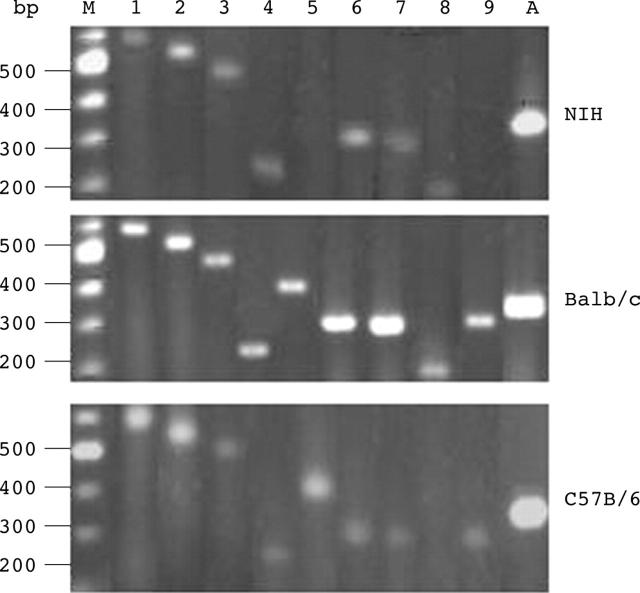
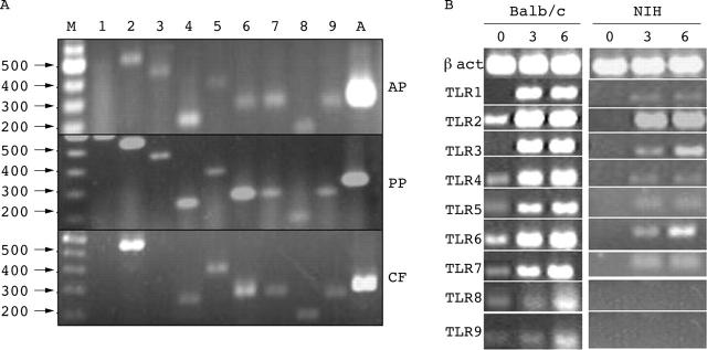
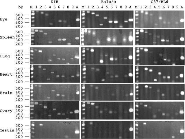
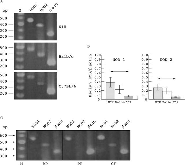
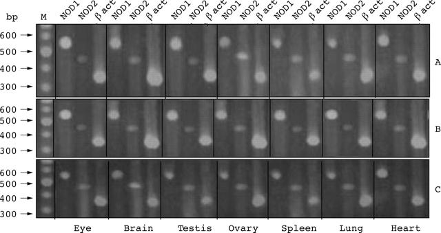
Methods: Semiquantitative RT-PCR was performed to look for TLR1-9 and NOD1 and NOD2 mRNA expressions in the whole eye, in the anterior (AP) and posterior (PP) portions of the eye, in corneal fibroblasts (CF) and in ovary, brain, testis, heart, lung, and spleen.

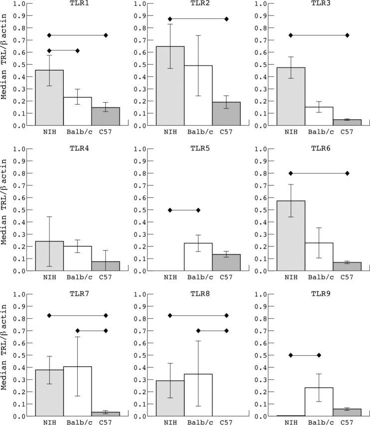
Results: All the TLR mRNAs were expressed in the whole eye of Balb/c mice. NIH and C57BL/6 did not express TLR9 and TLR8, respectively. NIH expressed higher levels of TLR1, 2, 3, and 6 than the other strains. C57BL/6 expressed the lowest levels of all TLRs. TLR9, 5, and 4 were the less expressed in all strains. All TLRs were expressed in Balb/c PP and TLR1 was not expressed in AP. In NIH and Balb/c CF the majority of TLRs were overexpressed with LPS. In testis, expression of most TLRs was absent. Non-immune privileged organs expressed most of the TLRs. All the organs expressed NOD1 and NOD2. In PP NOD2 was not expressed.

Conclusion: TLRs and NODs are expressed in the eye, and could have an important role in the innate immunity.


Figures

Figure 1
Figure 1
Expression of TLR mRNAs in the healthy eye of three mouse strains. RT-PCRs were performed in the eyes of NIH, Balb/c and C57BL/6 mice. Lane M corresponds to 100 bp molecular ladder; lanes 1–9 correspond to mRNA expression of TLRs1-9, respectively, and lane A to β actin expression.
Figure 2
Figure 2
Intensity of mRNA expression of TLRs in the eye of mice. The intensity of each band of figure 1 was measured in an Alpha Imager system. Each bar corresponds to the median value (SD) of the relation TLR/β actin mRNA expression in the whole eye of 10 NIH mice, five Balb/c mice, and five C57BL/6 mice, respectively. Horizontal lines show statistical difference (p<0.05) according to the Kruskal-Wallis test.
Figure 3
Figure 3
Expression of TLR mRNAs in different compartments of the eye. (A) RT-PCRs were performed in anterior portion (AP), posterior portion (PP), and corneal fibroblasts (CF) from a pool of 16 Balb/c mice eyes. In each gel image, lane M corresponds to 100 bp molecular ladder, lanes 1–9 correspond to mRNA expression of TLRs1-9, respectively, and lane A to β actin expression. (B) RT-PCRs were performed on TLR1-9 in corneal fibroblast of Balb/c and NIH mice treated at 0, 3, and 6 hours with LPS (10 μg/ml).
Figure 4
Figure 4
Expression of TLR mRNAs in non-immune privileged and immune privileged organs. RT-PCR was performed on whole eye, brain, testis, ovary, spleen, lung, and heart of NIH, Balb/c and C57BL/6 mice. In every gel image, lane M corresponds to 100 bp molecular ladder, lanes 1–9 correspond to mRNA expression of TLRs1-9, respectively, and lane A to β actin expression.
Figure 5
Figure 5
Expression of NOD1 and NOD2 mRNAs in the eye. (A) RT-PCR in whole eye of NIH, Balb/c, and C57BL/6 mice. Lane M corresponds to 100 bp molecular ladder. (B) Intensity of mRNA expression of NODs in the eye. Each bar shows the median (SD) of the relation NOD/β actin mRNA expression in the whole eye of 10 NIH, five Balb/c, and five C57BL/6 mice, respectively. Horizontal lines show statistical difference (p<0.05) by Kruskal-Wallis test. (C) mRNA expression of NODs in different eye compartments of Balb/c mice. AP, anterior portion; PP, posterior portion; CF, corneal fibroblasts.
Figure 6
Figure 6
Expression of NOD mRNA in non-immune privileged and immune privileged organs. RT-PCR from the whole eye, brain, testis, ovary, spleen, lung, and heart of NIH (A), Balb/c (B), and C57BL/6 (C) mice. Lane M: 100 bp molecular ladder.

References

    1. Wenkel H, Streilein JW. Analysis of immune deviation elicited by antigens injected into the subretinal space. Invest Ophthalmol Vis Sci 1998;39:1823–34. - PubMed
    1. Cousins SW, McCabe MM, Danielpour D, et al. Identification of transforming growth factor-beta as immunosuppressive factor in aqueous humor. Invest Ophthalmol Vis Sci 1991;32:2201–11. - PubMed
    1. Apte RS, Sinha D, Mayhew E, et al. Role of macrophage migration inhibitory factor in inhibiting NK cell activity and preserving immune privilege. J Immunol 1998;160:5693–6. - PubMed
    1. Hooper P, Bora NS, Kaplan HJ, et al. Inhibition of lymphocyte proliferation by resident ocular cells. Curr Eye Res 1991;10:363–72. - PubMed
    1. Ohta K, Wiggert B, Taylor AW, et al. Effects of experimental ocular inflammation on ocular immune privilege. Invest Ophtalmol Vis Sci 1999;40:2010–18. - PubMed

Publication types

MeSH terms