

Molecular genetic analysis of the nested Drosophila melanogaster lamin C gene
- PMID: 15965247
- PMCID: PMC1456510
- DOI: 10.1534/genetics.105.043208
Molecular genetic analysis of the nested Drosophila melanogaster lamin C gene
Abstract
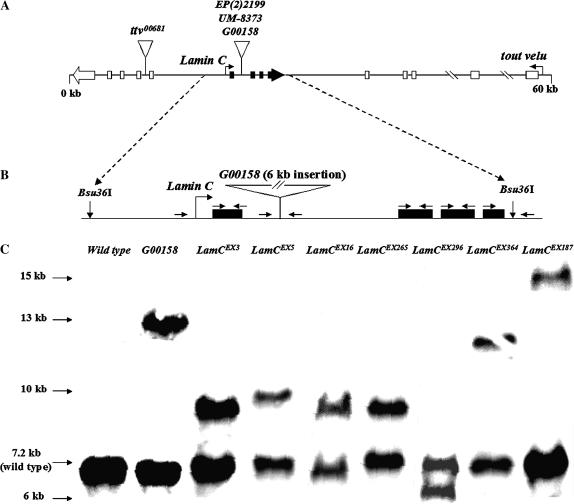
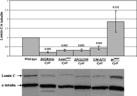
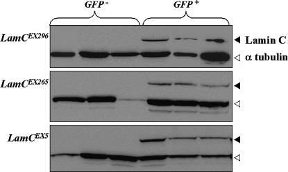
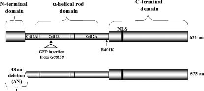
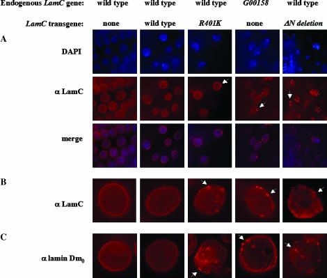
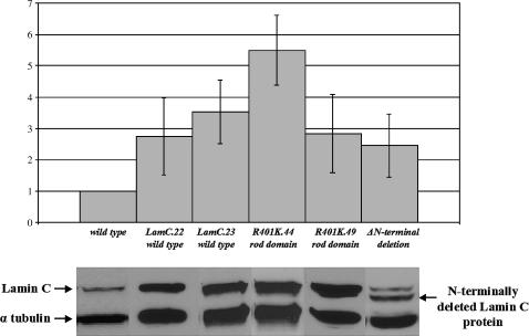
Lamins are intermediate filaments that line the inner surface of the nuclear envelope, providing structural support and making contacts with chromatin. There are two types of lamins, A- and B-types, which differ in structure and expression. Drosophila possesses both lamin types, encoded by the LamC (A-type) and lamin Dm0 (B-type) genes. LamC is nested within an intron of the essential gene ttv. We demonstrate that null mutations in LamC are lethal, and expression of a wild-type LamC transgene rescues lethality of LamC but not ttv mutants. Mutations in the human A-type lamin gene lead to diseases called laminopathies. To determine if Drosophila might serve as a useful model to study lamin biology and disease mechanisms, we generated transgenic flies expressing mutant LamC proteins modeled after human disease-causing lamins. These transgenic animals display a nuclear lamin aggregation phenotype remarkably similar to that observed when human mutant A-type lamins are expressed in mammalian cells. LamC aggregates also cause disorganization of lamin Dm0, indicating interdependence of both lamin types for proper lamina assembly. Taken together, these data provide the first detailed genetic analysis of the LamC gene and support using Drosophila as a model to study the role of lamins in disease.
Figures
Similar articles
-
Suppression of myopathic lamin mutations by muscle-specific activation of AMPK and modulation of downstream signaling.Hum Mol Genet. 2019 Feb 1;28(3):351-371. doi: 10.1093/hmg/ddy332. Hum Mol Genet. 2019. PMID: 30239736 Free PMC article.
-
Lamin C and chromatin organization in Drosophila.J Genet. 2010 Apr;89(1):37-49. doi: 10.1007/s12041-010-0009-y. J Genet. 2010. PMID: 20505245
-
A comparative study of Drosophila and human A-type lamins.PLoS One. 2009 Oct 26;4(10):e7564. doi: 10.1371/journal.pone.0007564. PLoS One. 2009. PMID: 19855837 Free PMC article.
-
Laminopathies: what can humans learn from fruit flies.Cell Mol Biol Lett. 2018 Jul 6;23:32. doi: 10.1186/s11658-018-0093-1. eCollection 2018. Cell Mol Biol Lett. 2018. PMID: 30002683 Free PMC article. Review.
-
Evolution of the lamin protein family: what introns can tell.Nucleus. 2012 Jan-Feb;3(1):44-59. doi: 10.4161/nucl.18927. Nucleus. 2012. PMID: 22156746 Review.
Cited by
-
The role of Drosophila Lamin C in muscle function and gene expression.Development. 2010 Sep;137(18):3067-77. doi: 10.1242/dev.048231. Epub 2010 Aug 11. Development. 2010. PMID: 20702563 Free PMC article.
-
Suppression of myopathic lamin mutations by muscle-specific activation of AMPK and modulation of downstream signaling.Hum Mol Genet. 2019 Feb 1;28(3):351-371. doi: 10.1093/hmg/ddy332. Hum Mol Genet. 2019. PMID: 30239736 Free PMC article.
-
Type-I prenyl protease function is required in the male germline of Drosophila melanogaster.G3 (Bethesda). 2012 Jun;2(6):629-42. doi: 10.1534/g3.112.002188. Epub 2012 Jun 1. G3 (Bethesda). 2012. PMID: 22690372 Free PMC article.
-
Myopathic lamin mutations cause reductive stress and activate the nrf2/keap-1 pathway.PLoS Genet. 2015 May 21;11(5):e1005231. doi: 10.1371/journal.pgen.1005231. eCollection 2015 May. PLoS Genet. 2015. PMID: 25996830 Free PMC article.
-
Age mosaic of gut epithelial cells prevents aging.Nat Commun. 2025 Jul 22;16(1):6734. doi: 10.1038/s41467-025-62043-y. Nat Commun. 2025. PMID: 40695820 Free PMC article.
References
-
- Adams, M. D., and J. J. Sekelsky, 2002. From sequence to phenotype: reverse genetics in Drosophila melanogaster. Nat. Rev. Genet. 3: 189–198. - PubMed
-
- Arimura, T., A. Helbling-Leclerc, C. Massart, S. Varnous, F. Niel et al., 2005. Mouse model carrying H222P-Lmna mutation develops muscular dystrophy and dilated cardiomyopathy similar to human striated muscle laminopathies. Hum. Mol. Genet. 14: 155–169. - PubMed
-
- Ashburner, M., 1989. Drosophila: A Laboratory Handbook. Cold Spring Harbor Laboratory Press, Cold Spring Harbor, NY.
-
- Bellaiche, Y., I. The and N. Perrimon, 1998. Tout-velu is a Drosophila homologue of the putative tumour suppressor EXT-1 and is needed for Hh diffusion. Nature 394: 85–88. - PubMed
-
- Bender, W., P. Spierer and D. S. Hogness, 1983. Chromosomal walking and jumping to isolate DNA from the Ace and rosy loci and the bithorax complex in Drosophila melanogaster. J. Mol. Biol. 168: 17–33. - PubMed
Publication types
MeSH terms
Substances
Grants and funding
LinkOut - more resources
Full Text Sources
Other Literature Sources
Molecular Biology Databases
