Comparative Study
. 2005 Oct;171(2):427-41.
doi: 10.1534/genetics.105.042861. Epub 2005 Jun 18.

Genetic instability induced by overexpression of DNA ligase I in budding yeast

Affiliations
Comparative Study

Genetic instability induced by overexpression of DNA ligase I in budding yeast

Jaichandar Subramanian et al. Genetics. 2005 Oct.

Abstract

Recombination and microsatellite mutation in humans contribute to disorders including cancer and trinucleotide repeat (TNR) disease. TNR expansions in wild-type yeast may arise by flap ligation during lagging-strand replication. Here we show that overexpression of DNA ligase I (CDC9) increases the rates of TNR expansion, of TNR contraction, and of mitotic recombination. Surprisingly, this effect is observed with catalytically inactive forms of Cdc9p protein, but only if they possess a functional PCNA-binding site. Furthermore, in vitro analysis indicates that the interaction of PCNA with Cdc9p and Rad27p (Fen1) is mutually exclusive. Together our genetic and biochemical analysis suggests that, although DNA ligase I seals DNA nicks during replication, repair, and recombination, higher than normal levels can yield genetic instability by disrupting the normal interplay of PCNA with other proteins such as Fen1.


Figures
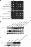
Figure 1.
Figure 1.
Characterization of CDC9 overexpression. (A) Complementation of a cdc9-2 mutant was performed by transforming LP2915-8D with a control vector (pRS316) or plasmids (pTW268 series; URA3) overexpressing different CDC9 alleles listed on the left. The transformants were resuspended in water and 10-fold dilutions were spotted on YPD at 30° (left) and 35° (right). Spot tests used three different dilutions (highest concentration on the left). (B) Western blot of Cdc9p. Cell extracts were obtained from BY4741 transformed with a control vector (pRS415; lane 1) or a plasmid overexpressing CDC9-NΔ23 (lane 2), GST-CDC9 (lane 3), GST-CDC9-NΔ23 (lane 4), GST-CDC9-NΔ60 (lane 5), GST-CDC9-K419A (lane 6), GST-CDC9-K598A (lane 7), or GST-CDC9-FF44,45AA (lane 8). Following 6% SDS-PAGE and transfer to a PVDF membrane, the membrane was cut and the proteins were probed with either a polyclonal anti-Cdc9p antiserum or a monoclonal anti-α-tubulin antibody. The top and middle rows represent the overexpressed ∼118-kDa GST-Cdc9p and the ∼85-kDa endogenous Cdc9p probed with the anti-Cdc9p antibody, respectively. The bottom row represents the ∼50-kDa α-tubulin protein (loading control). The numbers in parentheses represent the fold difference in the volume (intensity × square millimeters) of overexpressed GST-Cdc9p or its mutant versions compared to that of endogenous Cdc9p within the same lane. The endogenous Cdc9p in lane 2 cannot be distinguished from the overexpressed protein because they have the same molecular weight. (C) Purified PCNA (5 pmol of PCNA trimer) was incubated with glutathione sepharose beads liganded by 5 pmol of GST, GST-Cdc9p, or GST-Cdc9p-FF44,45AA. Proteins bound to GST beads (lane 2), GST-Cdc9p beads (lane 3), and GST-Cdc9p-FF44,45AA beads (lane 4) were separated by SDS-PAGE. GST-Cdc9p (top) and PCNA (bottom) were detected by immunblotting with antibodies to Cdc9p and yeast PCNA, respectively. Lane 1 represents 10% of the input PCNA used in the pull-down assay.
Figure 2.
Figure 2.
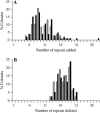
TNR size distributions estimated by single colony PCR analysis. (A) yJS1 colonies (with or without overexpression of CDC9) growing on media containing 5-FOA, but lacking histidine and leucine (expansion colonies), were used for PCR analysis. Data obtained from yJS1 colonies transformed with pRS415 or a plasmid overexpressing GST-CDC9-NΔ60 or GST-CDC9-FF44,45AA (collectively called PBM-WT colonies; shaded bars) were pooled and compared with the pooled data obtained from colonies transformed with a plasmid overexpressing GST-CDC9, GST-CDC9-NΔ23, GST-CDC9-K419A, or GST-CDC9-K598A (collectively called PBM-OE colonies; solid bars). The x-axis represents the number of repeats added to the original (CTG/CAG)25 and the y-axis represents the percentage of colonies carrying the expanded repeat size. For each of the two groups, PCR data from ∼100 individual colonies were pooled. (B) yJS3 colonies (with or without overexpression of CDC9) growing on media lacking leucine and uracil (contraction colonies) were used for PCR analysis. Data obtained from yJS3 colonies transformed with pRS415 or a plasmid overexpressing GST-CDC9-NΔ60 or GST-CDC9-FF44,45AA were pooled (PBM-WT colonies; shaded bars) and compared with the data pooled from colonies transformed with a plasmid overexpressing GST-CDC9, GST-CDC9-NΔ23, GST-CDC9-K419A, or GST-CDC9-K598A (PBM-OE colonies; solid bars). The x-axis represents the number of repeats deleted from the original (CTG/CAG)25 and the y-axis represents the percentage of colonies carrying the contracted repeat size. For each of the two groups, PCR data from ∼80 individual colonies were pooled.
Figure 3.
Figure 3.
Analysis of complexes formed by Rad27p, Cdc9p, and PCNA. To detect complexes formed among Rad27p, Cdc9p, and PCNA, we performed pull-down assays with Rad27p beads (referred to as B-R) as described in materials and methods. The presence (+) or absence (−) of any component (listed on the left) is indicated above. Lanes 1, 2, and 9 show purified PCNA, GST-Cdc9p, and GST-Cdc9p-FF44,45AA (GST-Cdc9p-M) proteins, respectively. These lanes contain 10% of the input proteins used in the pull-down assays. PCNA (5 pmol trimer) was incubated with Rad27p beads (lane 5). GST-Cdc9p or GST-Cdc9p-FF44,45AA (5 pmol of each) was incubated with Rad27p beads either alone (lanes, 6 and 11) or with PCNA (lanes 7 and 12, 5 pmol of trimer) as indicated. Similarly, a 5× molar excess (25 pmol) of either GST-Cdc9p (lane 8) or GST-Cdc9p-M (lane 13) was incubated with Rad27p beads in the presence of PCNA (5 pmol trimer). To control for background binding, PCNA (lane 3), GST-Cdc9p (lane 4), and GST-Cdc9p-FF44,45AA (lane 10) were each incubated with Ni-NTA agarose beads (B). Proteins eluted from the beads were separated by SDS-PAGE and detected by immunoblotting with Cdc9p (top), Rad27p (middle), and PCNA antibodies (bottom).
Figure 4.
Figure 4.
Recombination substrates. A detailed description of the constructs used for recombination assays can be found in the text. All constructs are represented with boxes and lines (not drawn to scale). Each box represents a gene listed on top. Similar boxes in the rest of the figure represent the same gene. The solid box indicates the location of the mutation. The broken lines represent the intervening sequence in the construct. (A) Substrate used for recombination assays at the LYS2 locus (not drawn to scale) contains a duplicated lys2 segment separated by sequences that include the HIS3 and URA3 genes. Pop-out recombination (left pathway) would result in the deletion of the intervening sequences and the formation of a functional LYS2 gene. Gene conversion (right pathway) would also produce a functional LYS2 gene without the loss of intervening sequence. (B) The pop-out recombination substrate used at the LEU2 locus contains a duplicate leu2-k allele, each with the same mutation (loss of a KpnI site). Therefore, pop-out recombination between the duplicates would still yield a mutated leu2-k allele. However, the ADE2 and URA3 genes present in the sequence between the two leu2-k fragments will be lost. (C) Recombination substrate used at the HIS3 locus for measuring gene conversion involves duplicate his3 fragments (nonfunctional). Gene conversion can generate a HIS3 fragment that does not carry both the mutations and, therefore, a functional gene.
Figure 5.
Figure 5.
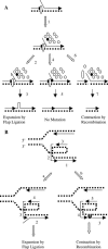
Model for TNR instability in wild-type yeast with and without CDC9 overexpression. (A) TNR instability induced by CDC9 overexpression. Once a flap is formed, overproduced DNA ligase I protein may out-compete Fen1 for binding to PCNA (1), which could increase the chance of TNR secondary structure formation (2) and lead to expansions by flap ligation (3). If Fen1 were able to cut the flap, mutation would be prevented (4, 5). Alternatively, delayed flap processing could lead to DSB formation (6) and a TNR contraction by recombination (7). Note that once the DSB is formed and the 5′-ends resected, the intermediate is identical regardless of repeat orientation (see text). The TNR sequence in the template strand is indicated by a broken line. Arrowheads mark the 3′-end of a nascent strand. The open ovals represent PCNA, the solid circles Fen1, and open circles DNA ligase I. (B) Orientation dependence of TNR instability in wild-type yeast (without CDC9 overexpression). This figure illustrates replication fork movement through a TNR sequence. Okazaki fragments (numbered) move counterclockwise as the replication fork advances to the left. (Path a) CTG flaps are formed in the lagging daughter strand and, following secondary structure formation, can cause expansions by flap ligation. (Path b) In the opposite orientation, secondary structure formation (represented as a U in the broken line) by CTG repeats in the single-stranded region (intermediate steps not shown) of the lagging-strand template leads to a DNA gap that can be repaired by recombination after a DNA break on the other strand and can produce a contraction mutation. Arrowheads correspond to the 3′-end of the daughter strands.

Similar articles

Cited by

References

    1. Ayyagari, R., X. V. Gomes, D. A. Gordenin and P. M. Burgers, 2003. Okazaki fragment maturation in yeast. I. Distribution of functions between FEN1 and DNA2. J. Biol. Chem. 278: 1618–1625. - PubMed
    1. Bae, S. H., and Y. S. Seo, 2000. Characterization of the enzymatic properties of the yeast dna2 helicase/endonuclease suggests a new model for Okazaki fragment processing. J. Biol. Chem. 275: 38022–38031. - PubMed
    1. Bae, S. H., K. H. Bae, J. A. Kim and Y. S. Seo, 2001. RPA governs endonuclease switching during processing of Okazaki fragments in eukaryotes. Nature 412: 456–461. - PubMed
    1. Barnes, D. E., L. H. Johnston, K. Kodama, A. E. Tomkinson, D. D. Lasko et al., 1990. Human DNA ligase I cDNA: cloning and functional expression in Saccharomyces cerevisiae. Proc. Natl. Acad. Sci. USA. 87: 6679–6683. - PMC - PubMed
    1. Bauer, G. A., and P. M. Burgers, 1988. The yeast analog of mammalian cyclin/proliferating-cell nuclear antigen interacts with mammalian DNA polymerase delta. Proc. Natl. Acad. Sci. USA. 85: 7506–7510. - PMC - PubMed

Publication types

MeSH terms